3U6Y

Crystal structure of ALBA2-DNA complex


Experimental Data Snapshot

  • Method: X-RAY DIFFRACTION
  • Resolution: 2.00 Å
  • R-Value Free: 0.282 
  • R-Value Work: 0.244 
  • R-Value Observed: 0.244 

wwPDB Validation   3D Report Full Report


This is version 1.2 of the entry. See complete history


Literature

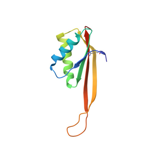
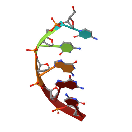
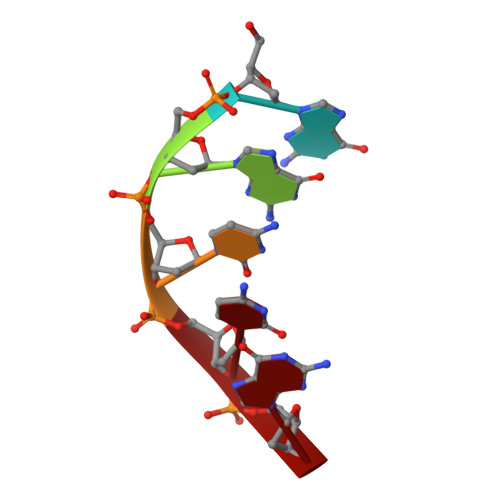
Crystal structure of archaeal chromatin protein Alba2-double-stranded DNA complex from Aeropyrum pernix K1.

Tanaka, T.Padavattan, S.Kumarevel, T.

(2012) J Biol Chem 287: 10394-10402

  • DOI: https://doi.org/10.1074/jbc.M112.343210
  • Primary Citation of Related Structures:  
    3U6Y

  • PubMed Abstract: 

    All thermophilic and hyperthermophilic archaea encode homologs of dimeric Alba (Sac10b) proteins that bind cooperatively at high density to DNA. Here, we report the 2.0 Å resolution crystal structure of an Alba2 (Ape10b2)-dsDNA complex from Aeropyrum pernix K1. A rectangular tube-like structure encompassing duplex DNA reveals the positively charged residues in the monomer-monomer interface of each dimer packing on either side of the bound dsDNA in successive minor grooves. The extended hairpin loop connecting strands β3 and β4 undergoes significant conformational changes upon DNA binding to accommodate the other Alba2 dimer during oligomerization. Mutational analysis of key interacting residues confirmed the specificity of Alba2-dsDNA interactions.


  • Organizational Affiliation

    RIKEN SPring-8 Center, Harima Institute, 1-1-1 Kouto, Sayo, Hyogo 679-5148, Japan.


Macromolecules

Find similar proteins by:  (by identity cutoff)  |  3D Structure
Entity ID: 1
MoleculeChains Sequence LengthOrganismDetailsImage
DNA/RNA-binding protein Alba 2A,
B [auth C]
102Aeropyrum pernix K1Mutation(s): 0 
Gene Names: albA2APE_1823
UniProt
Find proteins for Q9YAX2 (Aeropyrum pernix (strain ATCC 700893 / DSM 11879 / JCM 9820 / NBRC 100138 / K1))
Explore Q9YAX2 
Go to UniProtKB:  Q9YAX2
Entity Groups  
Sequence Clusters30% Identity50% Identity70% Identity90% Identity95% Identity100% Identity
UniProt GroupQ9YAX2
Sequence Annotations
Expand
  • Reference Sequence

Find similar nucleic acids by:  Sequence   |   3D Structure  

Entity ID: 2
MoleculeChains LengthOrganismImage
DNA (5'-D(*CP*CP*GP*GP*C)-3')C [auth D]5synthetic construct
Sequence Annotations
Expand
  • Reference Sequence

Find similar nucleic acids by:  Sequence   |   3D Structure  

Entity ID: 3
MoleculeChains LengthOrganismImage
DNA (5'-D(*GP*GP*CP*CP*G)-3')D [auth E]5synthetic construct
Sequence Annotations
Expand
  • Reference Sequence
Small Molecules
Ligands 1 Unique
IDChains Name / Formula / InChI Key2D Diagram3D Interactions
PO4
Query on PO4

Download Ideal Coordinates CCD File 
E [auth A]PHOSPHATE ION
O4 P
NBIIXXVUZAFLBC-UHFFFAOYSA-K
Experimental Data & Validation

Experimental Data

  • Method: X-RAY DIFFRACTION
  • Resolution: 2.00 Å
  • R-Value Free: 0.282 
  • R-Value Work: 0.244 
  • R-Value Observed: 0.244 
  • Space Group: P 21 21 21
Unit Cell:
Length ( Å )Angle ( ˚ )
a = 33.556α = 90
b = 74.29β = 90
c = 88.198γ = 90
Software Package:
Software NamePurpose
HKL-2000data collection
CNSrefinement
HKL-2000data reduction
HKL-2000data scaling
CNSphasing

Structure Validation

View Full Validation Report


Entry History 

Deposition Data

Revision History  (Full details and data files)

  • Version 1.0: 2012-02-22
    Type: Initial release
  • Version 1.1: 2020-01-01
    Changes: Database references, Source and taxonomy
  • Version 1.2: 2023-11-01
    Changes: Data collection, Database references, Derived calculations, Refinement description