3TRS

The crystal structure of aspergilloglutamic peptidase from Aspergillus niger


Experimental Data Snapshot

  • Method: X-RAY DIFFRACTION
  • Resolution: 1.60 Å
  • R-Value Free: 0.239 
  • R-Value Work: 0.208 
  • R-Value Observed: 0.210 

wwPDB Validation   3D Report Full Report


This is version 1.1 of the entry. See complete history


Literature

The crystal structure of an intermediate dimer of aspergilloglutamic peptidase that mimics the enzyme-activation product complex produced upon autoproteolysis.

Sasaki, H.Kubota, K.Lee, W.C.Ohtsuka, J.Kojima, M.Iwata, S.Nakagawa, A.Takahashi, K.Tanokura, M.

(2012) J Biochem 152: 45-52

  • DOI: https://doi.org/10.1093/jb/mvs050
  • Primary Citation of Related Structures:  
    3TRS

  • PubMed Abstract: 

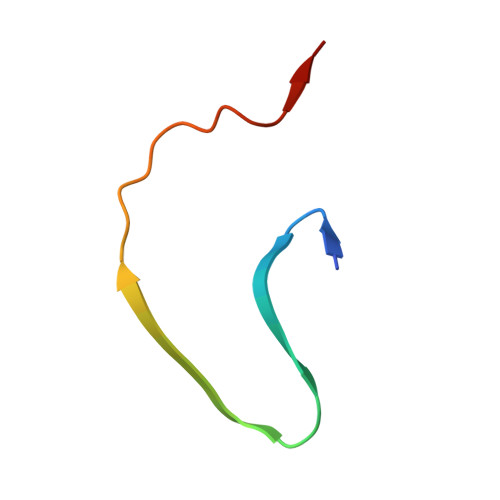
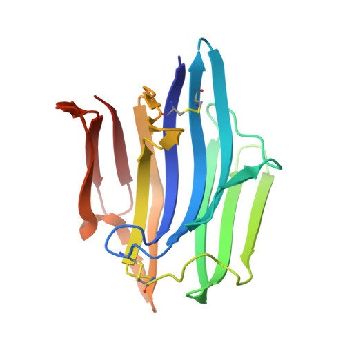
    Aspergilloglutamic peptidase from Aspergillus niger var. macrosporus (AGP) is one of the so-called pepstatin-insensitive acid endopeptidases, which are distinct from the well-studied aspartic peptidases. Among the known homologues of the glutamic peptidases, AGP is a unique two-chain enzyme with a light chain and a heavy chain bound non-covalently with each other, and thus is an interesting target for protein structure-function relationship studies. In this article, we report the crystal structure of a dimeric form of the enzyme at a resolution of 1.6 Å. This form has a unique structure in which the C-terminal region of the light chain of one of the molecules binds to the active site cleft of the other molecule like a part of a substrate. This form mimics the enzyme-activation product complex produced upon autoproteolysis, and provides a structural clue that could help to clarify the activation mechanism. This type of dimeric structure of a peptidase is here reported for the first time.


  • Organizational Affiliation

    Department of Biophysics and Biochemistry, Graduate School of Science, The University of Tokyo, 7-3-1 Hongo, Tokyo 113-0033, Japan.


Macromolecules
Find similar proteins by:  (by identity cutoff)  |  3D Structure
Entity ID: 1
MoleculeChains Sequence LengthOrganismDetailsImage
Aspergillopepsin-2 light chain
A, C
39Aspergillus niger var. macrosporusMutation(s): 0 
EC: 3.4.23.19
UniProt
Find proteins for P24665 (Aspergillus niger)
Explore P24665 
Go to UniProtKB:  P24665
Entity Groups  
Sequence Clusters30% Identity50% Identity70% Identity90% Identity95% Identity100% Identity
UniProt GroupP24665
Sequence Annotations
Expand
  • Reference Sequence
Find similar proteins by:  (by identity cutoff)  |  3D Structure
Entity ID: 2
MoleculeChains Sequence LengthOrganismDetailsImage
Aspergillopepsin-2 heavy chain
B, D
173Aspergillus niger var. macrosporusMutation(s): 0 
EC: 3.4.23.19
UniProt
Find proteins for P24665 (Aspergillus niger)
Explore P24665 
Go to UniProtKB:  P24665
Entity Groups  
Sequence Clusters30% Identity50% Identity70% Identity90% Identity95% Identity100% Identity
UniProt GroupP24665
Sequence Annotations
Expand
  • Reference Sequence
Small Molecules
Experimental Data & Validation

Experimental Data

  • Method: X-RAY DIFFRACTION
  • Resolution: 1.60 Å
  • R-Value Free: 0.239 
  • R-Value Work: 0.208 
  • R-Value Observed: 0.210 
  • Space Group: P 21 21 21
Unit Cell:
Length ( Å )Angle ( ˚ )
a = 60.692α = 90
b = 65.842β = 90
c = 77.391γ = 90
Software Package:
Software NamePurpose
MOLREPphasing
REFMACrefinement
DENZOdata reduction
SCALAdata scaling

Structure Validation

View Full Validation Report


Entry History 

Deposition Data

Revision History  (Full details and data files)

  • Version 1.0: 2012-08-22
    Type: Initial release
  • Version 1.1: 2023-11-01
    Changes: Data collection, Database references, Derived calculations, Refinement description