3TEQ

Crystal structure of SOAR domain


Experimental Data Snapshot

  • Method: X-RAY DIFFRACTION
  • Resolution: 1.90 Å
  • R-Value Free: 0.249 
  • R-Value Work: 0.219 
  • R-Value Observed: 0.219 

wwPDB Validation   3D Report Full Report


This is version 1.2 of the entry. See complete history


Literature

Structural and mechanistic insights into the activation of Stromal interaction molecule 1 (STIM1).

Yang, X.Jin, H.Cai, X.Li, S.Shen, Y.

(2012) Proc Natl Acad Sci U S A 109: 5657-5662

  • DOI: https://doi.org/10.1073/pnas.1118947109
  • Primary Citation of Related Structures:  
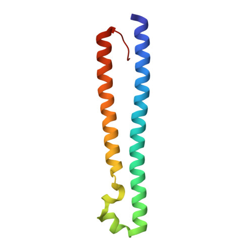
    3TEQ, 3TER

  • PubMed Abstract: 

    Calcium influx through the Ca(2+) release-activated Ca(2+) (CRAC) channel is an essential process in many types of cells. Upon store depletion, the calcium sensor in the endoplasmic reticulum, STIM1, activates Orai1, a CRAC channel in the plasma membrane. We have determined the structures of SOAR from Homo sapiens (hSOAR), which is part of STIM1 and is capable of constitutively activating Orai1, and the entire coiled coil region of STIM1 from Caenorhabditis elegans (ceSTIM1-CCR) in an inactive state. Our studies reveal that the formation of a SOAR dimer is necessary to activate the Orai1 channel. Mutations that disrupt SOAR dimerization or remove the cluster of positive residues abolish STIM1 activation of Orai1. We identified a possible inhibitory helix within the structure of ceSTIM1-CCR that tightly interacts with SOAR. Functional studies suggest that the inhibitory helix may keep the C-terminus of STIM1 in an inactive state. Our data allowed us to propose a model for STIM1 activation.


  • Organizational Affiliation

    State Key Laboratory of Medicinal Chemical Biology, College of Life Sciences, Nankai University, Tianjin 300071, China.


Macromolecules
Find similar proteins by:  (by identity cutoff)  |  3D Structure
Entity ID: 1
MoleculeChains Sequence LengthOrganismDetailsImage
Stromal interaction molecule 1
A, B, C, D
101Homo sapiensMutation(s): 3 
Gene Names: STIM1GOK
UniProt & NIH Common Fund Data Resources
Find proteins for Q13586 (Homo sapiens)
Explore Q13586 
Go to UniProtKB:  Q13586
PHAROS:  Q13586
GTEx:  ENSG00000167323 
Entity Groups  
Sequence Clusters30% Identity50% Identity70% Identity90% Identity95% Identity100% Identity
UniProt GroupQ13586
Sequence Annotations
Expand
  • Reference Sequence
Experimental Data & Validation

Experimental Data

  • Method: X-RAY DIFFRACTION
  • Resolution: 1.90 Å
  • R-Value Free: 0.249 
  • R-Value Work: 0.219 
  • R-Value Observed: 0.219 
  • Space Group: P 41 21 2
Unit Cell:
Length ( Å )Angle ( ˚ )
a = 90.584α = 90
b = 90.584β = 90
c = 104.919γ = 90
Software Package:
Software NamePurpose
MAR345dtbdata collection
PHENIXmodel building
CNSrefinement
HKL-2000data reduction
HKL-2000data scaling
PHENIXphasing

Structure Validation

View Full Validation Report


Entry History 

Deposition Data

Revision History  (Full details and data files)

  • Version 1.0: 2012-04-11
    Type: Initial release
  • Version 1.1: 2013-07-10
    Changes: Database references
  • Version 1.2: 2024-03-20
    Changes: Data collection, Database references, Derived calculations