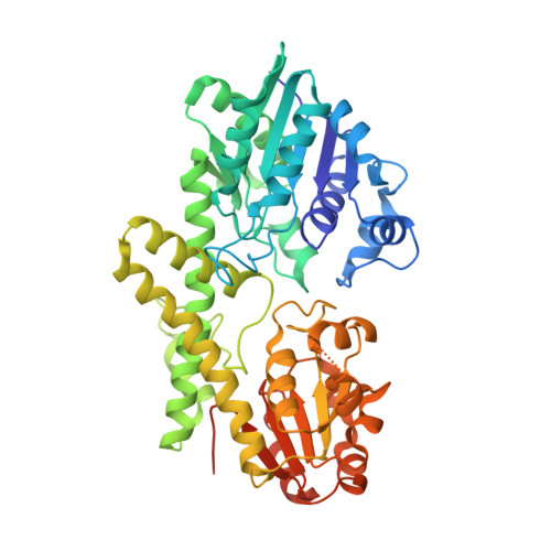
Structural Basis of Cooperativity in Human UDP-Glucose Dehydrogenase.
Rajakannan, V., Lee, H.S., Chong, S.H., Ryu, H.B., Bae, J.Y., Whang, E.Y., Huh, J.W., Cho, S.W., Kang, L.W., Choe, H., Robinson, R.C.(2011) PLoS One 6: e25226-e25226
- PubMed: 21984906
- DOI: https://doi.org/10.1371/journal.pone.0025226
- Primary Citation of Related Structures:
3TDK - PubMed Abstract:
UDP-glucose dehydrogenase (UGDH) is the sole enzyme that catalyzes the conversion of UDP-glucose to UDP-glucuronic acid. The product is used in xenobiotic glucuronidation in hepatocytes and in the production of proteoglycans that are involved in promoting normal cellular growth and migration. Overproduction of proteoglycans has been implicated in the progression of certain epithelial cancers, while inhibition of UGDH diminished tumor angiogenesis in vivo. A better understanding of the conformational changes occurring during the UGDH reaction cycle will pave the way for inhibitor design and potential cancer therapeutics. Previously, the substrate-bound of UGDH was determined to be a symmetrical hexamer and this regular symmetry is disrupted on binding the inhibitor, UDP-α-D-xylose. Here, we have solved an alternate crystal structure of human UGDH (hUGDH) in complex with UDP-glucose at 2.8 Å resolution. Surprisingly, the quaternary structure of this substrate-bound protein complex consists of the open homohexamer that was previously observed for inhibitor-bound hUGDH, indicating that this conformation is relevant for deciphering elements of the normal reaction cycle. In all subunits of the present open structure, Thr131 has translocated into the active site occupying the volume vacated by the absent active water and partially disordered NAD+ molecule. This conformation suggests a mechanism by which the enzyme may exchange NADH for NAD+ and repolarize the catalytic water bound to Asp280 while protecting the reaction intermediates. The structure also indicates how the subunits may communicate with each other through two reaction state sensors in this highly cooperative enzyme.
Organizational Affiliation:
Institute of Molecular and Cell Biology, Agency for Science, Technology and Research, Singapore, Singapore.


