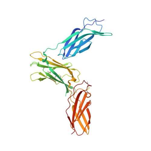
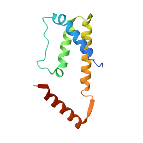
3QT2

Structure of a cytokine ligand-receptor complex


Experimental Data Snapshot

  • Method: X-RAY DIFFRACTION
  • Resolution: 2.55 Å
  • R-Value Free: 0.264 
  • R-Value Work: 0.207 
  • R-Value Observed: 0.210 

wwPDB Validation   3D Report Full Report


Ligand Structure Quality Assessment 


This is version 1.2 of the entry. See complete history


Literature

Structure Analysis of the IL-5 Ligand-Receptor Complex Reveals a Wrench-like Architecture for IL-5Ralpha.

Patino, E.Kotzsch, A.Saremba, S.Nickel, J.Schmitz, W.Sebald, W.Mueller, T.D.

(2011) Structure 19: 1864-1875

  • DOI: https://doi.org/10.1016/j.str.2011.08.015
  • Primary Citation of Related Structures:  
    3QT2

  • PubMed Abstract: 

    Interleukin-5 (IL-5) is the key mediator for the function of eosinophil granulocytes, whose deregulation is characteristic of hypereosinophilic diseases and presumably contributes to allergic asthma. IL-5 signaling involves two transmembrane receptors, IL-5Rα and the common β chain, which upon formation of the ternary complex activate the JAK/STAT signaling cascade. To investigate the mechanism underlying ligand-receptor recognition, we determined the structure of IL-5 bound to the extracellular domain of IL-5Rα. IL-5 makes contact with all three fibronectin III-like domains of IL-5Rα, with the receptor architecture resembling a wrench. Mutagenesis data provide evidence that this wrench-like architecture is likely preformed. The structure demonstrates that for steric reasons, homodimeric IL-5 can bind only one receptor molecule, even though two equivalent receptor-binding sites exist. In regard to strong efforts being made to develop IL-5 antagonists for treating asthma and hypereosinophilic diseases, the advances in molecular understanding provided by this structure are of greatest value.


  • Organizational Affiliation

    Lehrstuhl für Molekulare Pflanzenphysiologie und Biophysik, Julius-von-Sachs Institut der Universität Würzburg, Julius-von-Sachs Platz 2, D-97082 Würzburg, Germany.


Macromolecules
Find similar proteins by:  (by identity cutoff)  |  3D Structure
Entity ID: 1
MoleculeChains Sequence LengthOrganismDetailsImage
Interleukin-5 receptor subunit alphaA,
D [auth B]
317Homo sapiensMutation(s): 5 
Gene Names: IL5RIL5RA
UniProt & NIH Common Fund Data Resources
Find proteins for Q01344 (Homo sapiens)
Explore Q01344 
Go to UniProtKB:  Q01344
PHAROS:  Q01344
GTEx:  ENSG00000091181 
Entity Groups  
Sequence Clusters30% Identity50% Identity70% Identity90% Identity95% Identity100% Identity
UniProt GroupQ01344
Sequence Annotations
Expand
  • Reference Sequence
Find similar proteins by:  (by identity cutoff)  |  3D Structure
Entity ID: 2
MoleculeChains Sequence LengthOrganismDetailsImage
Interleukin-5B [auth C],
C [auth D],
E,
F
117Homo sapiensMutation(s): 0 
Gene Names: IL5
UniProt & NIH Common Fund Data Resources
Find proteins for P05113 (Homo sapiens)
Explore P05113 
Go to UniProtKB:  P05113
PHAROS:  P05113
GTEx:  ENSG00000113525 
Entity Groups  
Sequence Clusters30% Identity50% Identity70% Identity90% Identity95% Identity100% Identity
UniProt GroupP05113
Sequence Annotations
Expand
  • Reference Sequence
Experimental Data & Validation

Experimental Data

  • Method: X-RAY DIFFRACTION
  • Resolution: 2.55 Å
  • R-Value Free: 0.264 
  • R-Value Work: 0.207 
  • R-Value Observed: 0.210 
  • Space Group: P 1 21 1
Unit Cell:
Length ( Å )Angle ( ˚ )
a = 85.084α = 90
b = 61.633β = 99.49
c = 142.113γ = 90
Software Package:
Software NamePurpose
SHARPphasing
REFMACrefinement
CrystalCleardata reduction
CrystalCleardata scaling

Structure Validation

View Full Validation Report


Ligand Structure Quality Assessment 


Entry History 

Deposition Data

Revision History  (Full details and data files)

  • Version 1.0: 2012-02-01
    Type: Initial release
  • Version 1.1: 2017-11-08
    Changes: Refinement description
  • Version 1.2: 2020-07-29
    Type: Remediation
    Reason: Carbohydrate remediation
    Changes: Data collection, Database references, Derived calculations, Structure summary