3QRB

crystal structure of E-cadherin EC1-2 P5A P6A


Experimental Data Snapshot

  • Method: X-RAY DIFFRACTION
  • Resolution: 1.80 Å
  • R-Value Free: 0.178 
  • R-Value Work: 0.162 
  • R-Value Observed: 0.162 

wwPDB Validation   3D Report Full Report


This is version 1.3 of the entry. See complete history


Literature

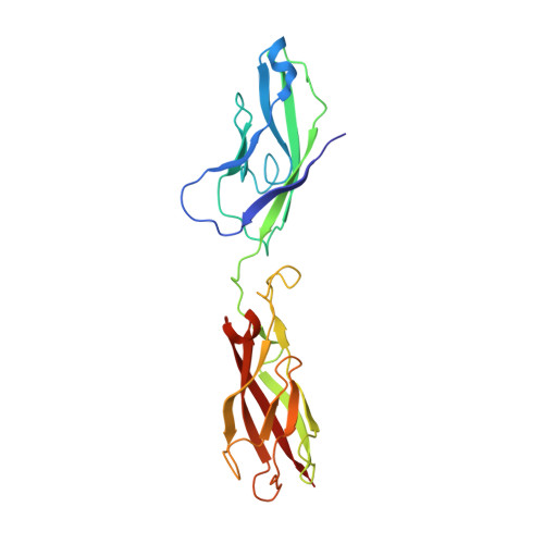
Molecular design principles underlying beta-strand swapping in the adhesive dimerization of cadherins.

Vendome, J.Posy, S.Jin, X.Bahna, F.Ahlsen, G.Shapiro, L.Honig, B.

(2011) Nat Struct Mol Biol 18: 693-700

  • DOI: https://doi.org/10.1038/nsmb.2051
  • Primary Citation of Related Structures:  
    3QRB

  • PubMed Abstract: 

    Cell adhesion by classical cadherins is mediated by dimerization of their EC1 domains through the 'swapping' of N-terminal β-strands. We use molecular simulations, measurements of binding affinities and X-ray crystallography to provide a detailed picture of the structural and energetic factors that control the adhesive dimerization of cadherins. We show that strand swapping in EC1 is driven by conformational strain in cadherin monomers that arises from the anchoring of their short N-terminal strand at one end by the conserved Trp2 and at the other by ligation to Ca(2+) ions. We also demonstrate that a conserved proline-proline motif functions to avoid the formation of an overly tight interface where affinity differences between different cadherins, crucial at the cellular level, are lost. We use these findings to design site-directed mutations that transform a monomeric EC2-EC3 domain cadherin construct into a strand-swapped dimer.


  • Organizational Affiliation

    Department of Biochemistry and Molecular Biophysics, Columbia University, New York, New York, USA. Center for Computational Biology and Bioinformatics, Columbia University, New York, New York, USA. Howard Hughes Medical Institute, Columbia University, New York, New York, USA.


Macromolecules
Find similar proteins by:  (by identity cutoff)  |  3D Structure
Entity ID: 1
MoleculeChains Sequence LengthOrganismDetailsImage
Cadherin-1
A, B
213Mus musculusMutation(s): 2 
Gene Names: Cdh1
UniProt & NIH Common Fund Data Resources
Find proteins for P09803 (Mus musculus)
Explore P09803 
Go to UniProtKB:  P09803
IMPC:  MGI:88354
Entity Groups  
Sequence Clusters30% Identity50% Identity70% Identity90% Identity95% Identity100% Identity
UniProt GroupP09803
Sequence Annotations
Expand
  • Reference Sequence
Small Molecules
Ligands 3 Unique
IDChains Name / Formula / InChI Key2D Diagram3D Interactions
PGE
Query on PGE

Download Ideal Coordinates CCD File 
H [auth A],
N [auth B]
TRIETHYLENE GLYCOL
C6 H14 O4
ZIBGPFATKBEMQZ-UHFFFAOYSA-N
EDO
Query on EDO

Download Ideal Coordinates CCD File 
F [auth A],
G [auth A],
L [auth B],
M [auth B]
1,2-ETHANEDIOL
C2 H6 O2
LYCAIKOWRPUZTN-UHFFFAOYSA-N
CA
Query on CA

Download Ideal Coordinates CCD File 
C [auth A]
D [auth A]
E [auth A]
I [auth B]
J [auth B]
C [auth A],
D [auth A],
E [auth A],
I [auth B],
J [auth B],
K [auth B]
CALCIUM ION
Ca
BHPQYMZQTOCNFJ-UHFFFAOYSA-N
Experimental Data & Validation

Experimental Data

  • Method: X-RAY DIFFRACTION
  • Resolution: 1.80 Å
  • R-Value Free: 0.178 
  • R-Value Work: 0.162 
  • R-Value Observed: 0.162 
  • Space Group: C 1 2 1
Unit Cell:
Length ( Å )Angle ( ˚ )
a = 111.185α = 90
b = 92.583β = 92.26
c = 79.465γ = 90
Software Package:
Software NamePurpose
ADSCdata collection
MOLREPphasing
REFMACrefinement
DENZOdata reduction
SCALEPACKdata scaling

Structure Validation

View Full Validation Report


Entry History 

Deposition Data

Revision History  (Full details and data files)

  • Version 1.0: 2011-05-18
    Type: Initial release
  • Version 1.1: 2011-07-13
    Changes: Version format compliance
  • Version 1.2: 2015-12-09
    Changes: Database references
  • Version 1.3: 2023-09-13
    Changes: Data collection, Database references, Derived calculations, Refinement description