Crystallographic, kinetic, and spectroscopic study of the first ligninolytic peroxidase presenting a catalytic tyrosine.
Miki, Y., Calvino, F.R., Pogni, R., Giansanti, S., Ruiz-Duenas, F.J., Martinez, M.J., Basosi, R., Romero, A., Martinez, A.T.(2011) J Biol Chem 286: 15525-15534
- PubMed: 21367853
- DOI: https://doi.org/10.1074/jbc.M111.220996
- Primary Citation of Related Structures:
3Q3U - PubMed Abstract:
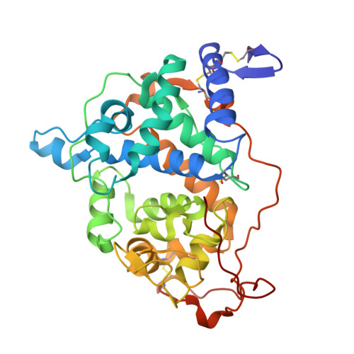
Trametes cervina lignin peroxidase (LiP) is a unique enzyme lacking the catalytic tryptophan strictly conserved in all other LiPs and versatile peroxidases (more than 30 sequences available). Recombinant T. cervina LiP and site-directed variants were investigated by crystallographic, kinetic, and spectroscopic techniques. The crystal structure shows three substrate oxidation site candidates involving His-170, Asp-146, and Tyr-181. Steady-state kinetics for oxidation of veratryl alcohol (the typical LiP substrate) by variants at the above three residues reveals a crucial role of Tyr-181 in LiP activity. Moreover, assays with ferrocytochrome c show that its ability to oxidize large molecules (a requisite property for oxidation of the lignin polymer) originates in Tyr-181. This residue is also involved in the oxidation of 1,4-dimethoxybenzene, a reaction initiated by the one-electron abstraction with formation of substrate cation radical, as described for the well known Phanerochaete chrysosporium LiP. Detailed spectroscopic and kinetic investigations, including low temperature EPR, show that the porphyrin radical in the two-electron activated T. cervina LiP is unstable and rapidly receives one electron from Tyr-181, forming a catalytic protein radical, which is identified as an H-bonded neutral tyrosyl radical. The crystal structure reveals a partially exposed location of Tyr-181, compatible with its catalytic role, and several neighbor residues probably contributing to catalysis: (i) by enabling substrate recognition by aromatic interactions; (ii) by acting as proton acceptor/donor from Tyr-181 or H-bonding the radical form; and (iii) by providing the acidic environment that would facilitate oxidation. This is the first structure-function study of the only ligninolytic peroxidase described to date that has a catalytic tyrosine.
Organizational Affiliation:
Centro de Investigaciones Biológicas, Consejo Superior de Investigaciones Científicas, Ramiro de Maeztu 9, E-28040 Madrid, Spain.