Biochemical studies and crystal structure determination of dihydrodipicolinate synthase from Pseudomonas aeruginosa
Kaur, N., Gautam, A., Kumar, S., Singh, A., Singh, N., Sharma, S., Sharma, R., Tewari, R., Singh, T.P.(2011) Int J Biol Macromol 48: 779-787
- PubMed: 21396954
- DOI: https://doi.org/10.1016/j.ijbiomac.2011.03.002
- Primary Citation of Related Structures:
3PS7, 3PUO - PubMed Abstract:
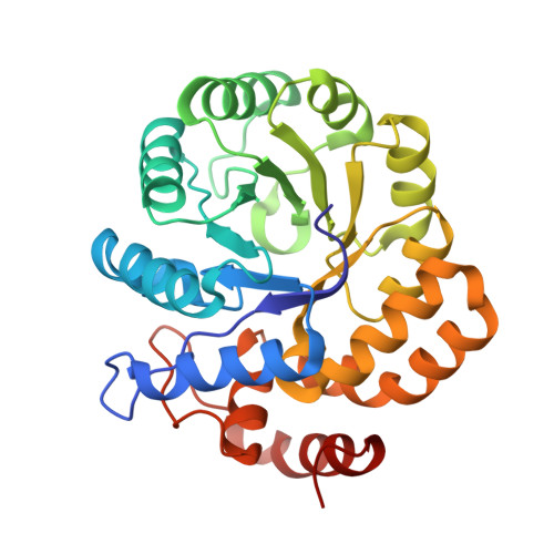
The intracellular enzyme dihydrodipicolinate synthase (DHDPS, E.C. 4.2.1.52) from Pseudomonas aeruginosa is a potential drug target because it is essential for the growth of bacteria while it is absent in humans. Therefore, in order to design new compounds using structure based approach for inhibiting the function of DHDPS from P. aeruginosa (Ps), we have cloned, characterized biochemically and biophysically and have determined its three-dimensional structure. The gene encoding DHDPS (dapA) was cloned in a vector pET-28c(+) and the recombinant protein was overexpressed in the Escherichia coli host. The K(m) values of the recombinant enzyme estimated for the substrates, pyruvate and (S)-aspartate-β-semialdehyde [(S)-ASA] were found to be 0.90±0.13 mM and 0.17±0.02 mM, respectively. The circular dichroism studies showed that the enzyme adopts a characteristic β/α conformation which is retained up to 65°C. The fluorescence data indicated the presence of exposed tryptophan residues in the enzyme. The three-dimensional structure determination showed that DHDPS forms a homodimer which is stabilized by several hydrogen bonds and van der Waals forces at the interface. The active site formed with residues Thr44, Tyr107 and Tyr133 is found to be stereochemically suitable for catalytic function. It may be noted that Tyr107 of the catalytic triad belongs to the partner molecule in the dimer. The structure of the complex of PsDHDPS with (S)-lysine determined at 2.65 Å resolution revealed the positions of three lysine molecules bound to the protein.
Organizational Affiliation:
Department of Biotechnology, Panjab University, Chandigarh, India.