3OBP

Anaerobic complex of urate oxidase with uric acid


Experimental Data Snapshot

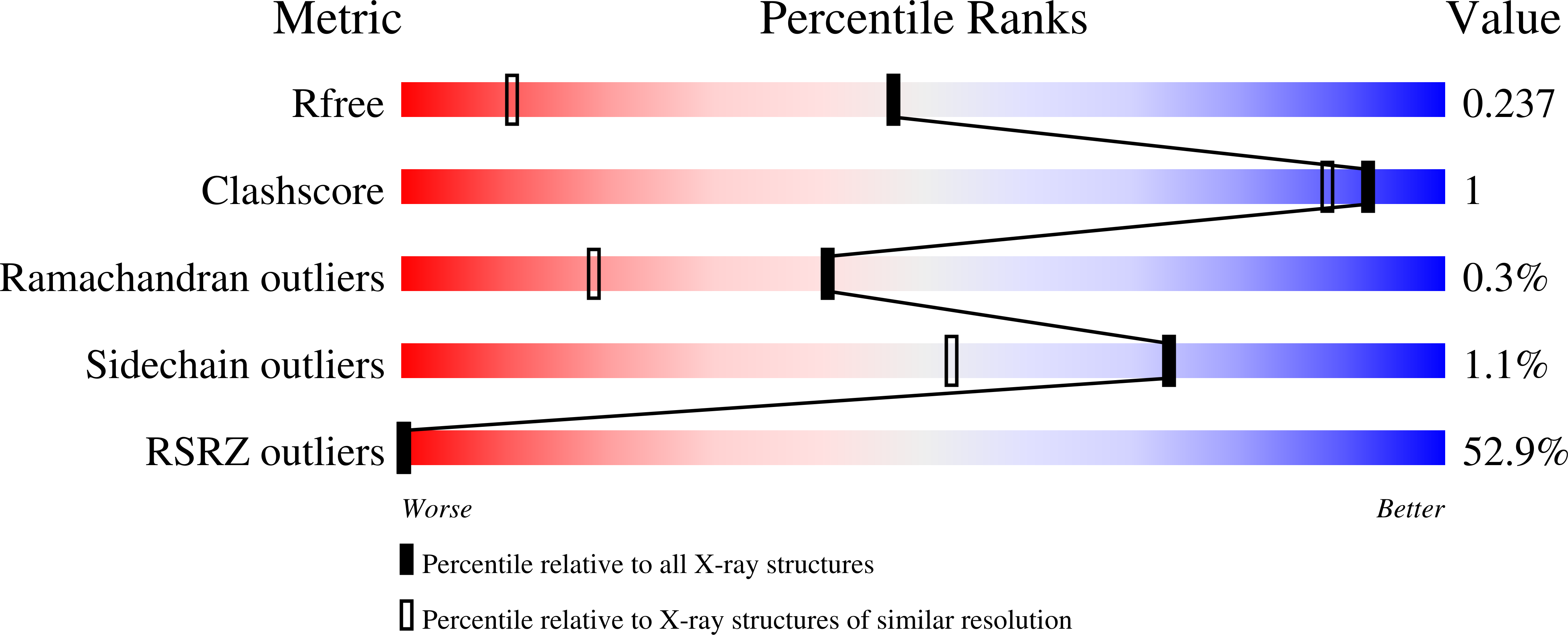
  • Method: X-RAY DIFFRACTION
  • Resolution: 1.50 Å
  • R-Value Free: 0.249 
  • R-Value Work: 0.220 
  • R-Value Observed: 0.228 

wwPDB Validation   3D Report Full Report


Ligand Structure Quality Assessment 


This is version 1.3 of the entry. See complete history


Literature

X-ray, ESR, and quantum mechanics studies unravel a spin well in the cofactor-less urate oxidase.

Gabison, L.Chopard, C.Colloc'h, N.Peyrot, F.Castro, B.Hajji, M.E.Altarsha, M.Monard, G.Chiadmi, M.Prange, T.

(2011) Proteins 79: 1964-1976

  • DOI: https://doi.org/10.1002/prot.23022
  • Primary Citation of Related Structures:  
    3OBP

  • PubMed Abstract: 

    Urate oxidase (EC 1.7.3.3 or UOX) catalyzes the conversion of uric acid using gaseous molecular oxygen to 5-hydroxyisourate and hydrogen peroxide in absence of any cofactor or transition metal. The catalytic mechanism was investigated using X-ray diffraction, electron spin resonance spectroscopy (ESR), and quantum mechanics calculations. The X-ray structure of the anaerobic enzyme-substrate complex gives credit to substrate activation before the dioxygen fixation in the peroxo hole, where incoming and outgoing reagents (dioxygen, water, and hydrogen peroxide molecules) are handled. ESR spectroscopy establishes the initial monoelectron activation of the substrate without the participation of dioxygen. In addition, both X-ray structure and quantum mechanic calculations promote a conserved base oxidative system as the main structural features in UOX that protonates/deprotonates and activate the substrate into the doublet state now able to satisfy the Wigner's spin selection rule for reaction with molecular oxygen in its triplet ground state.


  • Organizational Affiliation

    LCRB, UMR 8015 CNRS, Faculté de Pharmacie, Université Paris Descartes, 75270 Paris Cedex 06, France.


Macromolecules
Find similar proteins by:  (by identity cutoff)  |  3D Structure
Entity ID: 1
MoleculeChains Sequence LengthOrganismDetailsImage
Uricase302Aspergillus flavusMutation(s): 1 
Gene Names: uaZuox
EC: 1.7.3.3
UniProt
Find proteins for Q00511 (Aspergillus flavus)
Explore Q00511 
Go to UniProtKB:  Q00511
Entity Groups  
Sequence Clusters30% Identity50% Identity70% Identity90% Identity95% Identity100% Identity
UniProt GroupQ00511
Sequence Annotations
Expand
  • Reference Sequence
Experimental Data & Validation

Experimental Data

  • Method: X-RAY DIFFRACTION
  • Resolution: 1.50 Å
  • R-Value Free: 0.249 
  • R-Value Work: 0.220 
  • R-Value Observed: 0.228 
  • Space Group: I 2 2 2
Unit Cell:
Length ( Å )Angle ( ˚ )
a = 79.853α = 90
b = 96.29β = 90
c = 105.737γ = 90
Software Package:
Software NamePurpose
ADSCdata collection
SHELXL-97refinement
MOSFLMdata reduction
SCALAdata scaling
TRUNCATEdata scaling

Structure Validation

View Full Validation Report


Ligand Structure Quality Assessment 


Entry History 

Deposition Data

Revision History  (Full details and data files)

  • Version 1.0: 2011-06-22
    Type: Initial release
  • Version 1.1: 2011-07-13
    Changes: Version format compliance
  • Version 1.2: 2017-11-08
    Changes: Refinement description
  • Version 1.3: 2023-09-06
    Changes: Data collection, Database references, Derived calculations, Refinement description