3NX2

Enterobacter sp. Px6-4 Ferulic Acid Decarboxylase in complex with substrate analogues


Experimental Data Snapshot

  • Method: X-RAY DIFFRACTION
  • Resolution: 2.01 Å
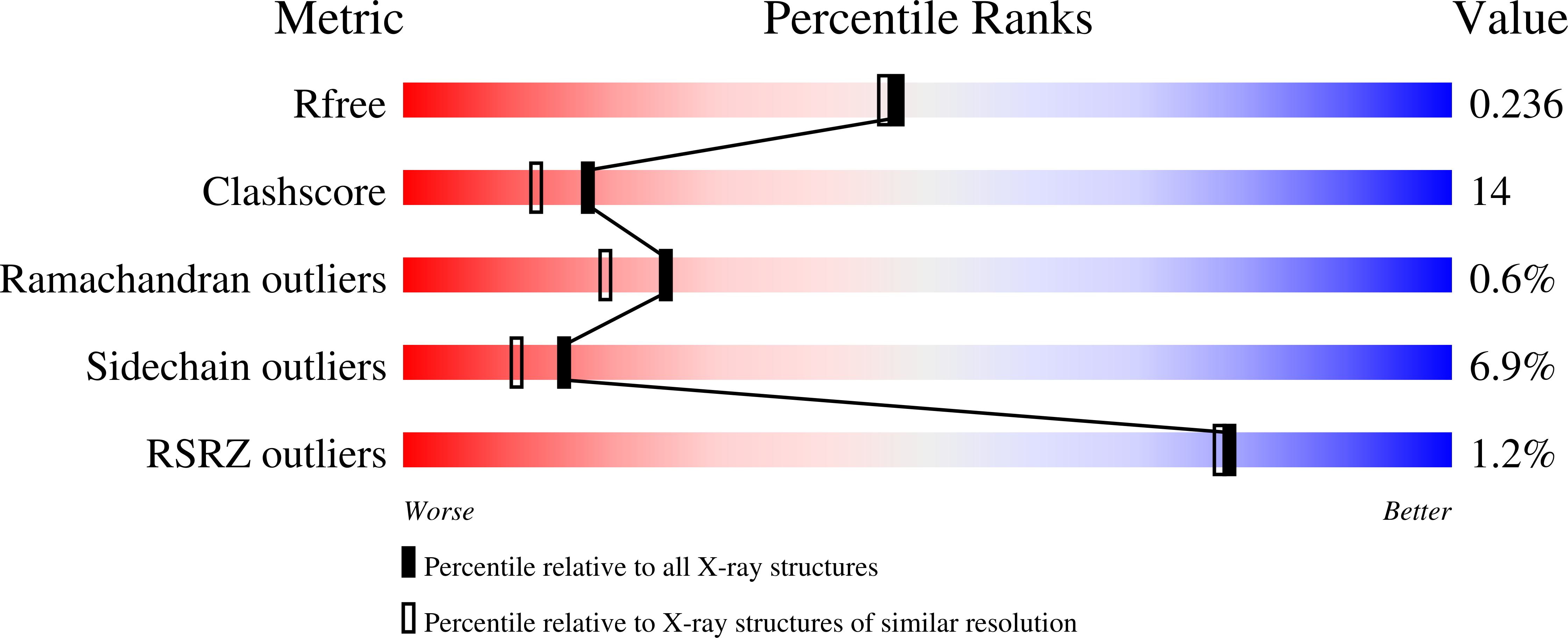
  • R-Value Free: 0.236 
  • R-Value Work: 0.189 
  • R-Value Observed: 0.191 

wwPDB Validation   3D Report Full Report


Ligand Structure Quality Assessment 


This is version 1.2 of the entry. See complete history


Literature

Structural Basis of Enzymatic Activity for the Ferulic Acid Decarboxylase (FADase) from Enterobacter sp. Px6-4

Gu, W.Yang, J.K.Lou, Z.Y.Liang, L.M.Sun, Y.Huang, J.W.Li, X.M.Cao, Y.Meng, Z.H.Zhang, K.-Q.

(2011) PLoS One 6: e16262-e16262

  • DOI: https://doi.org/10.1371/journal.pone.0016262
  • Primary Citation of Related Structures:  
    3NX1, 3NX2

  • PubMed Abstract: 

    Microbial ferulic acid decarboxylase (FADase) catalyzes the transformation of ferulic acid to 4-hydroxy-3-methoxystyrene (4-vinylguaiacol) via non-oxidative decarboxylation. Here we report the crystal structures of the Enterobacter sp. Px6-4 FADase and the enzyme in complex with substrate analogues. Our analyses revealed that FADase possessed a half-opened bottom β-barrel with the catalytic pocket located between the middle of the core β-barrel and the helical bottom. Its structure shared a high degree of similarity with members of the phenolic acid decarboxylase (PAD) superfamily. Structural analysis revealed that FADase catalyzed reactions by an "open-closed" mechanism involving a pocket of 8 × 8 × 15 Å dimension on the surface of the enzyme. The active pocket could directly contact the solvent and allow the substrate to enter when induced by substrate analogues. Site-directed mutagenesis showed that the E134A mutation decreased the enzyme activity by more than 60%, and Y21A and Y27A mutations abolished the enzyme activity completely. The combined structural and mutagenesis results suggest that during decarboxylation of ferulic acid by FADase, Trp25 and Tyr27 are required for the entering and proper orientation of the substrate while Glu134 and Asn23 participate in proton transfer.


  • Organizational Affiliation

    Laboratory for Conservation and Utilization of Bio-Resources, Key Laboratory for Microbial Resources of the Ministry of Education, Yunnan University, Kunming, China.


Macromolecules
Find similar proteins by:  (by identity cutoff)  |  3D Structure
Entity ID: 1
MoleculeChains Sequence LengthOrganismDetailsImage
Ferulic acid decarboxylase
A, B
168Enterobacter sp. Px6-4Mutation(s): 0 
EC: 4.1.1
UniProt
Find proteins for C6F3U5 (Enterobacter sp. Px6-4)
Explore C6F3U5 
Go to UniProtKB:  C6F3U5
Entity Groups  
Sequence Clusters30% Identity50% Identity70% Identity90% Identity95% Identity100% Identity
UniProt GroupC6F3U5
Sequence Annotations
Expand
  • Reference Sequence
Small Molecules
Ligands 1 Unique
IDChains Name / Formula / InChI Key2D Diagram3D Interactions
FER
Query on FER

Download Ideal Coordinates CCD File 
C [auth A]3-(4-HYDROXY-3-METHOXYPHENYL)-2-PROPENOIC ACID
C10 H10 O4
KSEBMYQBYZTDHS-HWKANZROSA-N
Experimental Data & Validation

Experimental Data

  • Method: X-RAY DIFFRACTION
  • Resolution: 2.01 Å
  • R-Value Free: 0.236 
  • R-Value Work: 0.189 
  • R-Value Observed: 0.191 
  • Space Group: P 1 21 1
Unit Cell:
Length ( Å )Angle ( ˚ )
a = 43.334α = 90
b = 88.74β = 102.3
c = 49.017γ = 90
Software Package:
Software NamePurpose
MAR345dtbdata collection
PHASERphasing
REFMACrefinement
HKL-2000data reduction
HKL-2000data scaling

Structure Validation

View Full Validation Report


Ligand Structure Quality Assessment 


Entry History 

Deposition Data

Revision History  (Full details and data files)

  • Version 1.0: 2011-02-16
    Type: Initial release
  • Version 1.1: 2011-07-13
    Changes: Version format compliance
  • Version 1.2: 2023-11-01
    Changes: Data collection, Database references, Derived calculations, Refinement description