


Crystal Structure of Glucose-1-phosphatase (AgpE) from Enterobacter cloacae
Grishkovskaya, I., Herter, T., Wessner, H., Borriss, R., Hoehne, W.To be published.
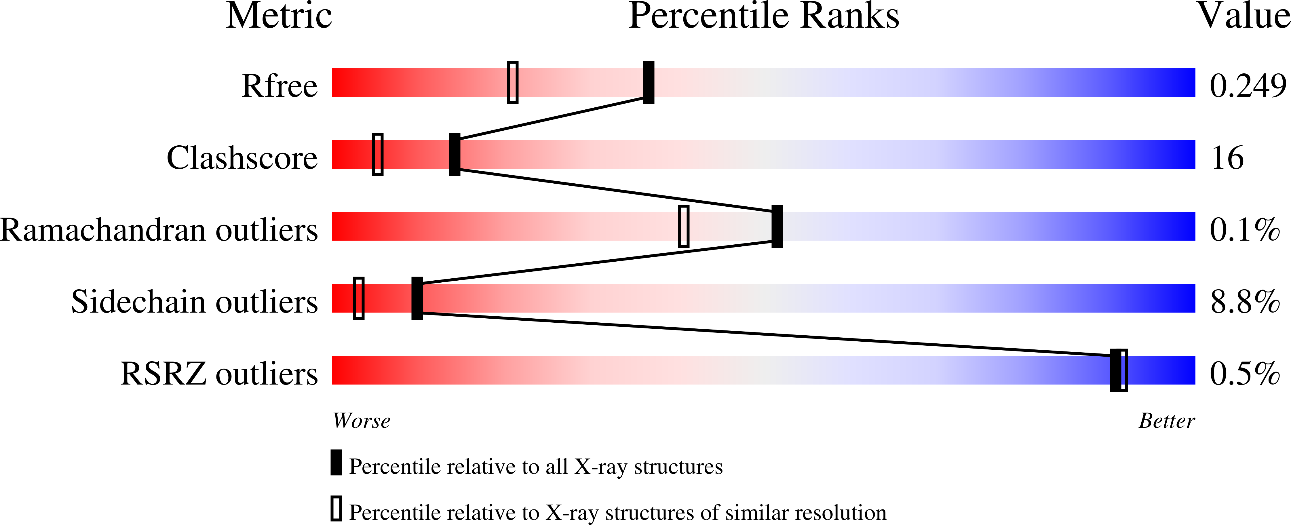
Experimental Data Snapshot
Entity ID: 1 | |||||
|---|---|---|---|---|---|
| Molecule | Chains | Sequence Length | Organism | Details | Image |
| Acid glucose-1-phosphate phosphatase | 398 | Enterobacter cloacae | Mutation(s): 1 Gene Names: agp | 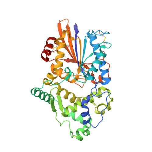 | |
UniProt | |||||
Find proteins for Q6EV19 (Enterobacter cloacae) Explore Q6EV19 Go to UniProtKB: Q6EV19 | |||||
Entity Groups | |||||
| Sequence Clusters | 30% Identity50% Identity70% Identity90% Identity95% Identity100% Identity | ||||
| UniProt Group | Q6EV19 | ||||
Sequence AnnotationsExpand | |||||
| |||||
| Ligands 3 Unique | |||||
|---|---|---|---|---|---|
| ID | Chains | Name / Formula / InChI Key | 2D Diagram | 3D Interactions | |
| IHP Query on IHP | C [auth A], E [auth B] | INOSITOL HEXAKISPHOSPHATE C6 H18 O24 P6 IMQLKJBTEOYOSI-GPIVLXJGSA-N |  | ||
| PO4 Query on PO4 | D [auth A] | PHOSPHATE ION O4 P NBIIXXVUZAFLBC-UHFFFAOYSA-K |  | ||
| CA Query on CA | F [auth B] | CALCIUM ION Ca BHPQYMZQTOCNFJ-UHFFFAOYSA-N |  | ||
| Length ( Å ) | Angle ( ˚ ) |
|---|---|
| a = 151.079 | α = 90 |
| b = 151.079 | β = 90 |
| c = 86.644 | γ = 120 |
| Software Name | Purpose |
|---|---|
| HKL-2000 | data collection |
| MOLREP | phasing |
| REFMAC | refinement |
| HKL-2000 | data reduction |
| SCALA | data scaling |
RCSB PDB (citation) is hosted by
RCSB PDB is a member of the

