3MLG

2ouf-2x, a designed knotted protein


Experimental Data Snapshot

  • Method: X-RAY DIFFRACTION
  • Resolution: 2.29 Å
  • R-Value Free: 0.278 
  • R-Value Work: 0.248 
  • R-Value Observed: 0.250 

wwPDB Validation   3D Report Full Report


This is version 1.4 of the entry. See complete history


Literature

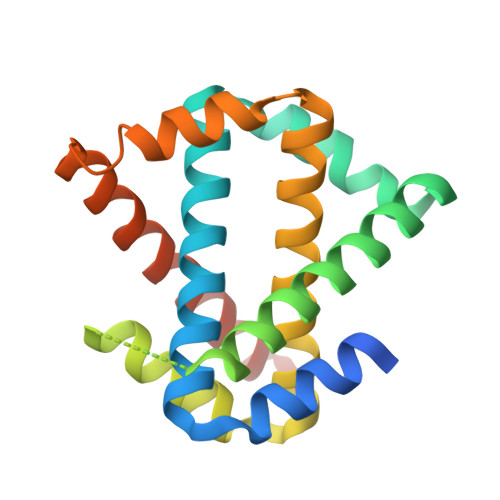
Structure and folding of a designed knotted protein.

King, N.P.Jacobitz, A.W.Sawaya, M.R.Goldschmidt, L.Yeates, T.O.

(2010) Proc Natl Acad Sci U S A 107: 20732-20737

  • DOI: https://doi.org/10.1073/pnas.1007602107
  • Primary Citation of Related Structures:  
    3MLG, 3MLI

  • PubMed Abstract: 

    A very small number of natural proteins have folded configurations in which the polypeptide backbone is knotted. Relatively little is known about the folding energy landscapes of such proteins, or how they have evolved. We explore those questions here by designing a unique knotted protein structure. Biophysical characterization and X-ray crystal structure determination show that the designed protein folds to the intended configuration, tying itself in a knot in the process, and that it folds reversibly. The protein folds to its native, knotted configuration approximately 20 times more slowly than a control protein, which was designed to have a similar tertiary structure but to be unknotted. Preliminary kinetic experiments suggest a complicated folding mechanism, providing opportunities for further characterization. The findings illustrate a situation where a protein is able to successfully traverse a complex folding energy landscape, though the amino acid sequence of the protein has not been subjected to evolutionary pressure for that ability. The success of the design strategy--connecting two monomers of an intertwined homodimer into a single protein chain--supports a model for evolution of knotted structures via gene duplication.


  • Organizational Affiliation

    Department of Chemistry and Biochemistry, University of California, Los Angeles, CA 90095-1569, USA.


Macromolecules
Find similar proteins by:  (by identity cutoff)  |  3D Structure
Entity ID: 1
MoleculeChains Sequence LengthOrganismDetailsImage
2X chimera of Helicobacter pylori protein HP0242
A, B
189Helicobacter pylori 26695Mutation(s): 0 
Gene Names: HP_0242
UniProt
Find proteins for O25025 (Helicobacter pylori (strain ATCC 700392 / 26695))
Explore O25025 
Go to UniProtKB:  O25025
Entity Groups  
Sequence Clusters30% Identity50% Identity70% Identity90% Identity95% Identity100% Identity
UniProt GroupO25025
Sequence Annotations
Expand
  • Reference Sequence
Experimental Data & Validation

Experimental Data

  • Method: X-RAY DIFFRACTION
  • Resolution: 2.29 Å
  • R-Value Free: 0.278 
  • R-Value Work: 0.248 
  • R-Value Observed: 0.250 
  • Space Group: P 21 21 21
Unit Cell:
Length ( Å )Angle ( ˚ )
a = 44.942α = 90
b = 75.39β = 90
c = 118.573γ = 90
Software Package:
Software NamePurpose
HKL-2000data processing
PHASERphasing
BUSTER-TNTrefinement
PDB_EXTRACTdata extraction
ADSCdata collection
DENZOdata reduction
SCALEPACKdata scaling

Structure Validation

View Full Validation Report


Entry History 

Deposition Data

Revision History  (Full details and data files)

  • Version 1.0: 2010-05-12
    Type: Initial release
  • Version 1.1: 2011-07-13
    Changes: Version format compliance
  • Version 1.2: 2014-04-09
    Changes: Source and taxonomy
  • Version 1.3: 2017-07-26
    Changes: Database references, Refinement description, Source and taxonomy, Structure summary
  • Version 1.4: 2023-09-06
    Changes: Data collection, Database references, Refinement description