3LDI

Crystal structure of aprotinin in complex with sucrose octasulfate: unusual interactions and implication for heparin binding


Experimental Data Snapshot

  • Method: X-RAY DIFFRACTION
  • Resolution: 2.20 Å
  • R-Value Free: 0.234 
  • R-Value Work: 0.206 
  • R-Value Observed: 0.207 

wwPDB Validation   3D Report Full Report


This is version 1.2 of the entry. See complete history


Literature

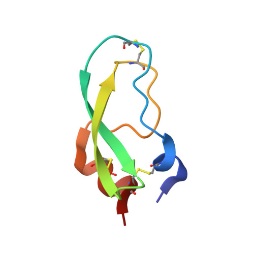
Crystal structures of aprotinin and its complex with sucrose octasulfate reveal multiple modes of interactions with implications for heparin binding

Yang, I.S.Kim, T.G.Park, B.S.Cho, K.J.Lee, J.-H.Park, Y.Kim, K.H.

(2010) Biochem Biophys Res Commun 397: 429-435

  • DOI: https://doi.org/10.1016/j.bbrc.2010.05.113
  • Primary Citation of Related Structures:  
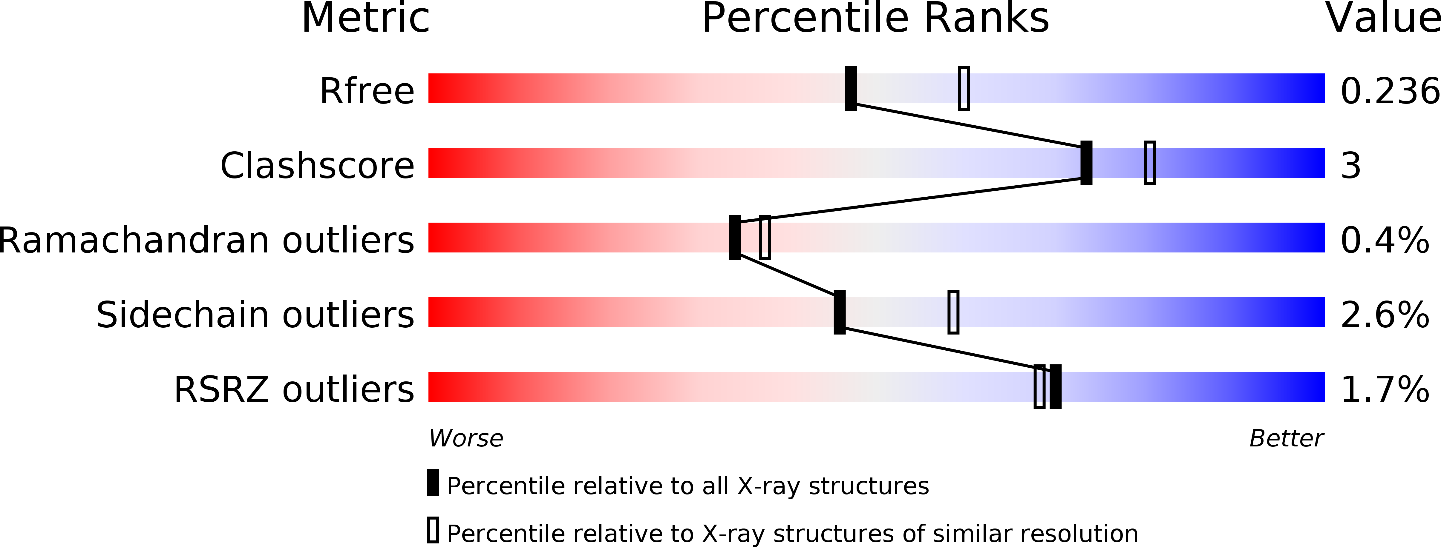
    3LDI, 3LDJ, 3LDM

  • PubMed Abstract: 

    The crystal structures of aprotinin and its complex with sucrose octasulfate (SOS), a polysulfated heparin analog, were determined at 1.7-2.6A resolutions. Aprotinin is monomeric in solution, which associates into a decamer at high salt concentrations. Sulfate ions serve to neutralize the basic amino acid residues of aprotinin to stabilize the decameric aprotinin. Whereas SOS interacts with heparin binding proteins at 1:1 molar ratio, SOS was surprisingly found to induce strong agglutination of aprotinins. Five molecules of aprotinin interact with one molecule of the sulfated sugar, which is stabilized by electrostatic interactions between the positively charged residues of aprotinin and sulfate groups of SOS. The multiple binding modes of SOS with five individual aprotinin molecules may represent the diverse patterns of potential heparin binding to aprotinin, reflecting the interactions of densely packed protein molecules along the heparin polymer.


  • Organizational Affiliation

    Department of Biotechnology & Bioinformatics, Korea University, Chungnam 339-700, Republic of Korea.


Macromolecules
Find similar proteins by:  (by identity cutoff)  |  3D Structure
Entity ID: 1
MoleculeChains Sequence LengthOrganismDetailsImage
Pancreatic trypsin inhibitor
A, B, C, D, E
58Bos taurusMutation(s): 0 
UniProt
Find proteins for P00974 (Bos taurus)
Explore P00974 
Go to UniProtKB:  P00974
Entity Groups  
Sequence Clusters30% Identity50% Identity70% Identity90% Identity95% Identity100% Identity
UniProt GroupP00974
Sequence Annotations
Expand
  • Reference Sequence
Small Molecules
Ligands 3 Unique
IDChains Name / Formula / InChI Key2D Diagram3D Interactions
HG
Query on HG

Download Ideal Coordinates CCD File 
DA [auth E],
J [auth A],
P [auth B],
T [auth C],
Y [auth D]
MERCURY (II) ION
Hg
BQPIGGFYSBELGY-UHFFFAOYSA-N
SO4
Query on SO4

Download Ideal Coordinates CCD File 
AA [auth E]
BA [auth E]
CA [auth E]
F [auth A]
G [auth A]
AA [auth E],
BA [auth E],
CA [auth E],
F [auth A],
G [auth A],
H [auth A],
I [auth A],
K [auth B],
L [auth B],
M [auth B],
N [auth B],
Q [auth C],
R [auth C],
U [auth D],
V [auth D],
W [auth D],
X [auth D],
Z [auth E]
SULFATE ION
O4 S
QAOWNCQODCNURD-UHFFFAOYSA-L
GOL
Query on GOL

Download Ideal Coordinates CCD File 
O [auth B],
S [auth C]
GLYCEROL
C3 H8 O3
PEDCQBHIVMGVHV-UHFFFAOYSA-N
Experimental Data & Validation

Experimental Data

  • Method: X-RAY DIFFRACTION
  • Resolution: 2.20 Å
  • R-Value Free: 0.234 
  • R-Value Work: 0.206 
  • R-Value Observed: 0.207 
  • Space Group: P 63 2 2
Unit Cell:
Length ( Å )Angle ( ˚ )
a = 119.08α = 90
b = 119.08β = 90
c = 110.794γ = 120
Software Package:
Software NamePurpose
MOLREPphasing
REFMACrefinement
PDB_EXTRACTdata extraction
HKL-2000data collection
HKL-2000data reduction
HKL-2000data scaling

Structure Validation

View Full Validation Report


Entry History 

Deposition Data

Revision History  (Full details and data files)

  • Version 1.0: 2010-06-23
    Type: Initial release
  • Version 1.1: 2011-07-13
    Changes: Version format compliance
  • Version 1.2: 2014-02-12
    Changes: Database references