Structure, mechanism, and substrate profile for Sco3058: the closest bacterial homologue to human renal dipeptidase .
Cummings, J.A., Nguyen, T.T., Fedorov, A.A., Kolb, P., Xu, C., Fedorov, E.V., Shoichet, B.K., Barondeau, D.P., Almo, S.C., Raushel, F.M.(2010) Biochemistry 49: 611-622
- PubMed: 20000809
- DOI: https://doi.org/10.1021/bi901935y
- Primary Citation of Related Structures:
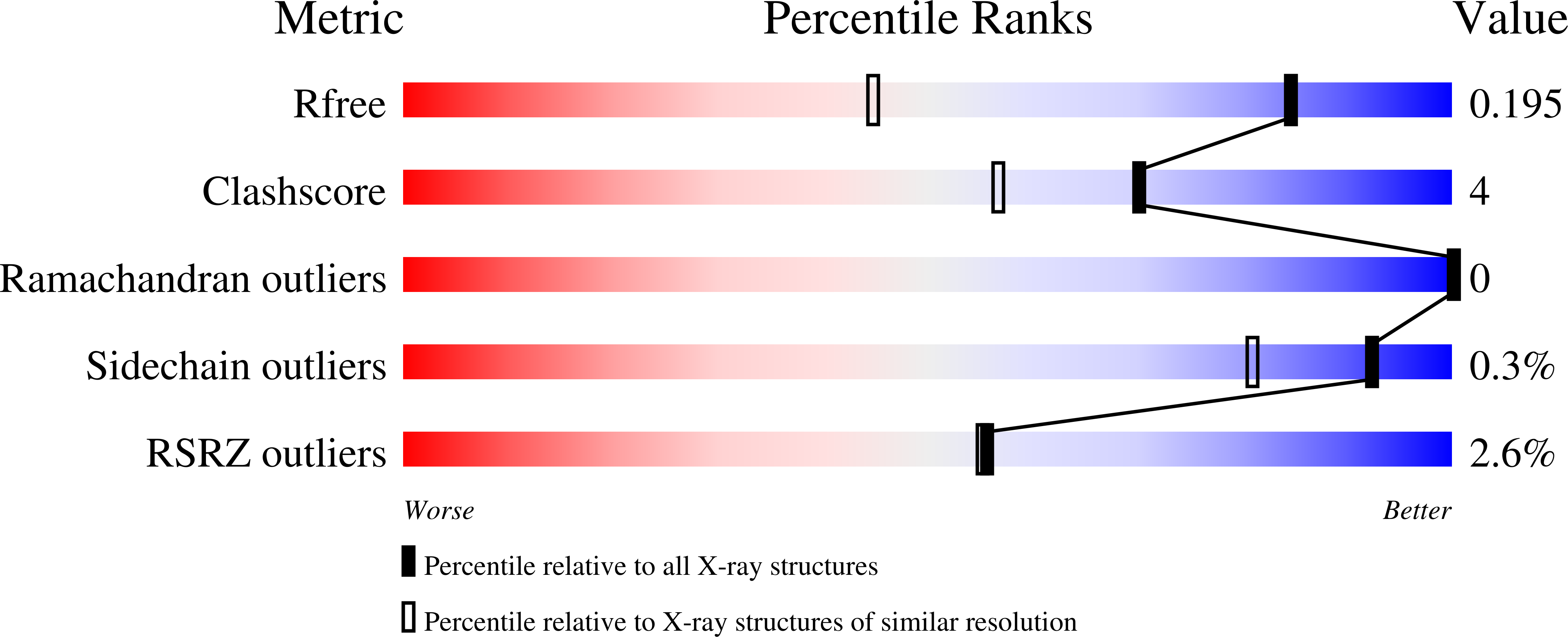
3ID7, 3ITC, 3K5X - PubMed Abstract:
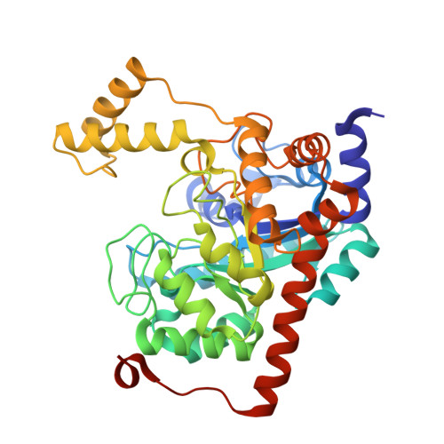
Human renal dipeptidase, an enzyme associated with glutathione metabolism and the hydrolysis of beta-lactams, is similar in sequence to a cluster of approximately 400 microbial proteins currently annotated as nonspecific dipeptidases within the amidohydrolase superfamily. The closest homologue to the human renal dipeptidase from a fully sequenced microbe is Sco3058 from Streptomyces coelicolor. Dipeptide substrates of Sco3058 were identified by screening a comprehensive series of l-Xaa-l-Xaa, l-Xaa-d-Xaa, and d-Xaa-l-Xaa dipeptide libraries. The substrate specificity profile shows that Sco3058 hydrolyzes a broad range of dipeptides with a marked preference for an l-amino acid at the N-terminus and a d-amino acid at the C-terminus. The best substrate identified was l-Arg-d-Asp (k(cat)/K(m) = 7.6 x 10(5) M(-1) s(-1)). The three-dimensional structure of Sco3058 was determined in the absence and presence of the inhibitors citrate and a phosphinate mimic of l-Ala-d-Asp. The enzyme folds as a (beta/alpha)(8) barrel, and two zinc ions are bound in the active site. Site-directed mutagenesis was used to probe the importance of specific residues that have direct interactions with the substrate analogues in the active site (Asp-22, His-150, Arg-223, and Asp-320). The solvent viscosity and kinetic effects of D(2)O indicate that substrate binding is relatively sticky and that proton transfers do not occurr during the rate-limiting step. A bell-shaped pH-rate profile for k(cat) and k(cat)/K(m) indicated that one group needs to be deprotonated and a second group must be protonated for optimal turnover. Computational docking of high-energy intermediate forms of l/d-Ala-l/d-Ala to the three-dimensional structure of Sco3058 identified the structural determinants for the stereochemical preferences for substrate binding and turnover.
Organizational Affiliation:
Department of Chemistry, P.O. Box 30012, Texas A&M University, College Station, Texas 77843, USA.