3JVE

Crystal Structure of the Sixth BRCT Domain of TopBP1


Experimental Data Snapshot

  • Method: X-RAY DIFFRACTION
  • Resolution: 1.34 Å
  • R-Value Free: 0.173 
  • R-Value Work: 0.158 
  • R-Value Observed: 0.159 

wwPDB Validation   3D Report Full Report


This is version 1.3 of the entry. See complete history


Literature

Insights from the crystal structure of the sixth BRCT domain of topoisomerase IIbeta binding protein 1.

Leung, C.C.Kellogg, E.Kuhnert, A.Hanel, F.Baker, D.Glover, J.N.

(2010) Protein Sci 19: 162-167

  • DOI: https://doi.org/10.1002/pro.290
  • Primary Citation of Related Structures:  
    3JVE

  • PubMed Abstract: 

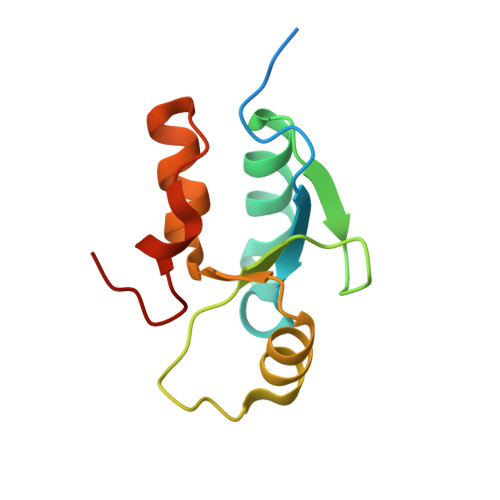
    Topoisomerase IIbeta binding protein 1 (TopBP1) is a major player in the DNA damage response and interacts with a number of protein partners via its eight BRCA1 carboxy-terminal (BRCT) domains. In particular, the sixth BRCT domain of TopBP1 has been implicated in binding to the phosphorylated transcription factor, E2F1, and poly(ADP-ribose) polymerase 1 (PARP-1), where the latter interaction is responsible for the poly(ADP-ribosyl)ation of TopBP1. To gain a better understanding of the nature of TopBP1 BRCT6 interactions, we solved the crystal structure of BRCT6 to 1.34 A. The crystal structure reveals a degenerate phospho-peptide binding pocket and lacks conserved hydrophobic residues involved in packing of tandem BRCT repeats, which, together with results from phospho-peptide binding studies, strongly suggest that TopBP1 BRCT6 independently does not function as a phospho-peptide binding domain. We further provide insight into poly(ADP-ribose) binding and sites of potential modification by PARP-1.


  • Organizational Affiliation

    Department of Biochemistry, University of Alberta, Edmonton, Alberta, Canada T6G 2H7.


Macromolecules
Find similar proteins by:  (by identity cutoff)  |  3D Structure
Entity ID: 1
MoleculeChains Sequence LengthOrganismDetailsImage
DNA topoisomerase 2-binding protein 1109Homo sapiensMutation(s): 0 
Gene Names: TOPBP1KIAA0259
UniProt & NIH Common Fund Data Resources
Find proteins for Q92547 (Homo sapiens)
Explore Q92547 
Go to UniProtKB:  Q92547
PHAROS:  Q92547
GTEx:  ENSG00000163781 
Entity Groups  
Sequence Clusters30% Identity50% Identity70% Identity90% Identity95% Identity100% Identity
UniProt GroupQ92547
Sequence Annotations
Expand
  • Reference Sequence
Experimental Data & Validation

Experimental Data

  • Method: X-RAY DIFFRACTION
  • Resolution: 1.34 Å
  • R-Value Free: 0.173 
  • R-Value Work: 0.158 
  • R-Value Observed: 0.159 
  • Space Group: P 21 21 21
Unit Cell:
Length ( Å )Angle ( ˚ )
a = 35.767α = 90
b = 51.824β = 90
c = 62.086γ = 90
Software Package:
Software NamePurpose
DENZOdata reduction
SCALEPACKdata scaling
PHASERphasing
REFMACrefinement
PDB_EXTRACTdata extraction
HKL-2000data collection

Structure Validation

View Full Validation Report


Entry History 

Deposition Data

Revision History  (Full details and data files)

  • Version 1.0: 2010-01-19
    Type: Initial release
  • Version 1.1: 2011-07-13
    Changes: Version format compliance
  • Version 1.2: 2017-11-01
    Changes: Refinement description
  • Version 1.3: 2024-02-21
    Changes: Data collection, Database references