3J04

EM structure of the heavy meromyosin subfragment of Chick smooth muscle Myosin with regulatory light chain in phosphorylated state


Experimental Data Snapshot

  • Method: ELECTRON MICROSCOPY
  • Resolution: 20.0 Å
  • Aggregation State: 2D ARRAY 
  • Reconstruction Method: CRYSTALLOGRAPHY 

wwPDB Validation   3D Report Full Report


This is version 1.3 of the entry. See complete history


Literature

Phosphorylated smooth muscle heavy meromyosin shows an open conformation linked to activation.

Baumann, B.A.Taylor, D.W.Huang, Z.Tama, F.Fagnant, P.M.Trybus, K.M.Taylor, K.A.

(2012) J Mol Biol 415: 274-287

  • DOI: https://doi.org/10.1016/j.jmb.2011.10.047
  • Primary Citation of Related Structures:  
    3J04

  • PubMed Abstract: 

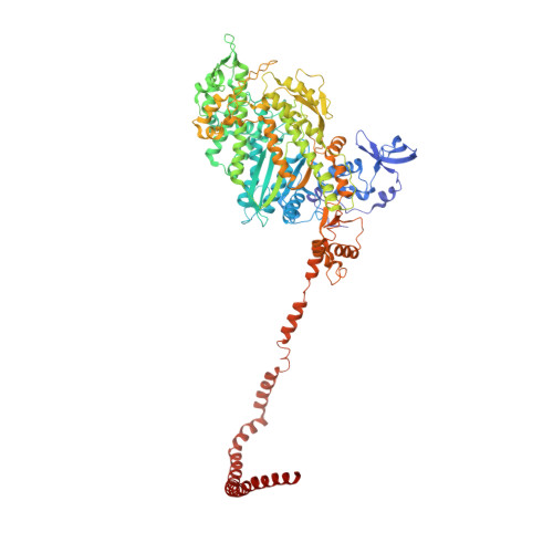
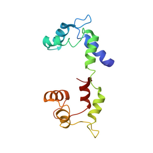
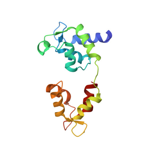
    Smooth muscle myosin and smooth muscle heavy meromyosin (smHMM) are activated by regulatory light chain phosphorylation, but the mechanism remains unclear. Dephosphorylated, inactive smHMM assumes a closed conformation with asymmetric intramolecular head-head interactions between motor domains. The "free head" can bind to actin, but the actin binding interface of the "blocked head" is involved in interactions with the free head. We report here a three-dimensional structure for phosphorylated, active smHMM obtained using electron crystallography of two-dimensional arrays. Head-head interactions of phosphorylated smHMM resemble those found in the dephosphorylated state but occur between different molecules, not within the same molecule. The light chain binding domain structure of phosphorylated smHMM differs markedly from that of the "blocked" head of dephosphorylated smHMM. We hypothesize that regulatory light chain phosphorylation opens the inhibited conformation primarily by its effect on the blocked head. Singly phosphorylated smHMM is not compatible with the closed conformation if the blocked head is phosphorylated. This concept has implications for the extent of myosin activation at low levels of phosphorylation in smooth muscle.


  • Organizational Affiliation

    Institute of Molecular Biophysics, Florida State University, Tallahassee, FL 32306, USA.


Macromolecules
Find similar proteins by:  (by identity cutoff)  |  3D Structure
Entity ID: 1
MoleculeChains Sequence LengthOrganismDetailsImage
Myosin-11
A, D
909Gallus gallusMutation(s): 0 
UniProt
Find proteins for P10587 (Gallus gallus)
Explore P10587 
Go to UniProtKB:  P10587
Entity Groups  
Sequence Clusters30% Identity50% Identity70% Identity90% Identity95% Identity100% Identity
UniProt GroupP10587
Sequence Annotations
Expand
  • Reference Sequence
Find similar proteins by:  (by identity cutoff)  |  3D Structure
Entity ID: 2
MoleculeChains Sequence LengthOrganismDetailsImage
Myosin regulatory light chain 2, smooth muscle major isoform
B, E
143Gallus gallusMutation(s): 0 
UniProt
Find proteins for P02612 (Gallus gallus)
Explore P02612 
Go to UniProtKB:  P02612
Entity Groups  
Sequence Clusters30% Identity50% Identity70% Identity90% Identity95% Identity100% Identity
UniProt GroupP02612
Sequence Annotations
Expand
  • Reference Sequence
Find similar proteins by:  (by identity cutoff)  |  3D Structure
Entity ID: 3
MoleculeChains Sequence LengthOrganismDetailsImage
Myosin light polypeptide 6
C, F
148Gallus gallusMutation(s): 0 
UniProt
Find proteins for P02607 (Gallus gallus)
Explore P02607 
Go to UniProtKB:  P02607
Entity Groups  
Sequence Clusters30% Identity50% Identity70% Identity90% Identity95% Identity100% Identity
UniProt GroupP02607
Sequence Annotations
Expand
  • Reference Sequence
Experimental Data & Validation

Experimental Data

  • Method: ELECTRON MICROSCOPY
  • Resolution: 20.0 Å
  • Aggregation State: 2D ARRAY 
  • Reconstruction Method: CRYSTALLOGRAPHY 

Structure Validation

View Full Validation Report


Entry History 

Deposition Data

Revision History  (Full details and data files)

  • Version 1.0: 2011-11-16
    Type: Initial release
  • Version 1.1: 2011-11-30
    Changes: Database references
  • Version 1.2: 2012-02-01
    Changes: Database references
  • Version 1.3: 2024-02-21
    Changes: Author supporting evidence, Data collection, Database references