3F1S

Crystal structure of Protein Z complexed with protein Z-dependent inhibitor


Experimental Data Snapshot

  • Method: X-RAY DIFFRACTION
  • Resolution: 2.30 Å
  • R-Value Free: 0.271 
  • R-Value Work: 0.224 
  • R-Value Observed: 0.227 

wwPDB Validation   3D Report Full Report


Ligand Structure Quality Assessment 


This is version 1.3 of the entry. See complete history


Literature

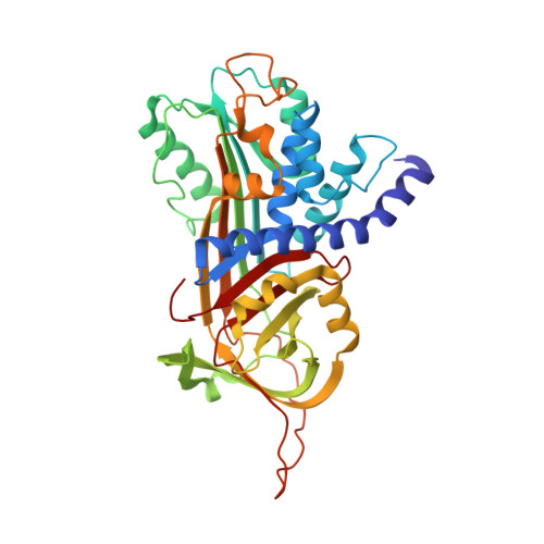
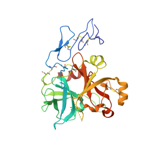
Crystal structure of protein Z-dependent inhibitor complex shows how protein Z functions as a cofactor in the membrane inhibition of factor X.

Wei, Z.Yan, Y.Carrell, R.W.Zhou, A.

(2009) Blood 

  • DOI: https://doi.org/10.1182/blood-2009-04-210021
  • Primary Citation of Related Structures:  
    3F1S

  • PubMed Abstract: 

    Protein Z (PZ) binds to PZ-dependent inhibitor (ZPI) and accelerates the inhibition of the coagulation protease, activated factor X (FXa), in the presence of phospholipids and Ca2+. A 2.3A resolution crystal structure of PZ complexed with ZPI shows that ZPI is a typical serine protease inhibitor and that PZ has a serine protease fold with distorted oxyanion hole and S1 pocket. The 2 molecules bind with fully complementary surfaces spanning over 2400A(2) and involving extensive ionic and hydrophobic interactions. ZPI has an unusual shutter region with a negatively charged residue buried within the hydrophobic core of the molecule. This unique Asp(213) is critical in maintaining the balanced metastability required for optimal protease inhibition, especially when PZ is bound, with its replacement with Asn resulting in increased thermal stability, but decreased efficiency of protease inhibition. The structure of ZPI shows negatively and positively charged surfaces on top of the molecule, in keeping with mutagenesis studies in this work indicating exosite interactions with FXa when it docks on top of ZPI. As modeled in this study, the gamma-carboxy-glutamic acid-containing domains of PZ and FXa enable them to bind to the same phospholipid surfaces on platelet and other membranes, with optimal proximity for the inhibition of FXa by the complexed ZPI.


  • Organizational Affiliation

    Department of Haematology, University of Cambridge, Cambridge Institute for Medical Research, Cambridge, United Kingdom.


Macromolecules
Find similar proteins by:  (by identity cutoff)  |  3D Structure
Entity ID: 1
MoleculeChains Sequence LengthOrganismDetailsImage
Protein Z-dependent protease inhibitor385Homo sapiensMutation(s): 0 
Gene Names: SERPINA10UNQ707/PRO1358ZPI
UniProt & NIH Common Fund Data Resources
Find proteins for Q9UK55 (Homo sapiens)
Explore Q9UK55 
Go to UniProtKB:  Q9UK55
PHAROS:  Q9UK55
GTEx:  ENSG00000140093 
Entity Groups  
Sequence Clusters30% Identity50% Identity70% Identity90% Identity95% Identity100% Identity
UniProt GroupQ9UK55
Sequence Annotations
Expand
  • Reference Sequence
Find similar proteins by:  (by identity cutoff)  |  3D Structure
Entity ID: 2
MoleculeChains Sequence LengthOrganismDetailsImage
Vitamin K-dependent protein Z283Homo sapiensMutation(s): 0 
Gene Names: PROZPZ
UniProt & NIH Common Fund Data Resources
Find proteins for P22891 (Homo sapiens)
Explore P22891 
Go to UniProtKB:  P22891
PHAROS:  P22891
GTEx:  ENSG00000126231 
Entity Groups  
Sequence Clusters30% Identity50% Identity70% Identity90% Identity95% Identity100% Identity
UniProt GroupP22891
Sequence Annotations
Expand
  • Reference Sequence
Experimental Data & Validation

Experimental Data

  • Method: X-RAY DIFFRACTION
  • Resolution: 2.30 Å
  • R-Value Free: 0.271 
  • R-Value Work: 0.224 
  • R-Value Observed: 0.227 
  • Space Group: P 21 21 21
Unit Cell:
Length ( Å )Angle ( ˚ )
a = 68.562α = 90
b = 96.514β = 90
c = 117.077γ = 90
Software Package:
Software NamePurpose
MOSFLMdata reduction
PHASERphasing
REFMACrefinement
PDB_EXTRACTdata extraction

Structure Validation

View Full Validation Report


Ligand Structure Quality Assessment 


Entry History 

Deposition Data

  • Released Date: 2009-06-30 
  • Deposition Author(s): Zhou, A.

Revision History  (Full details and data files)

  • Version 1.0: 2009-06-30
    Type: Initial release
  • Version 1.1: 2011-07-13
    Changes: Advisory, Version format compliance
  • Version 1.2: 2020-07-29
    Type: Remediation
    Reason: Carbohydrate remediation
    Changes: Data collection, Database references, Derived calculations, Structure summary
  • Version 1.3: 2023-12-27
    Changes: Data collection, Database references, Structure summary