3EZI

Crystal Structure of the E. coli Histidine Kinase NarX Sensor Domain without Ligand


Experimental Data Snapshot

  • Method: X-RAY DIFFRACTION
  • Resolution: 1.70 Å
  • R-Value Free: 0.239 
  • R-Value Work: 0.189 

wwPDB Validation   3D Report Full Report


This is version 1.4 of the entry. See complete history


Literature

Structural Analysis of Ligand Stimulation of the Histidine Kinase NarX.

Cheung, J.Hendrickson, W.A.

(2009) Structure 17: 190-201

  • DOI: https://doi.org/10.1016/j.str.2008.12.013
  • Primary Citation of Related Structures:  
    3EZH, 3EZI

  • PubMed Abstract: 

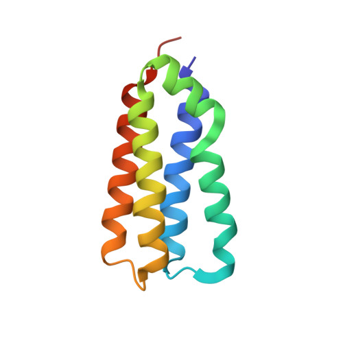
    Histidine kinase receptors are a large family of membrane-spanning proteins found in many prokaryotes and some eukaryotes. They are a part of two-component signal transduction systems, which each comprise a sensor kinase and a response regulator and are involved with the regulation of many cellular processes. NarX is a histidine kinase receptor that responds to nitrate and nitrite to effect regulation of anaerobic respiration in various bacteria. We present high-resolution X-ray crystal structures of the periplasmic sensor domain from Escherichia coli NarX in a complex with nitrate and in the apo state. Our analysis reveals that nitrate-binding induces conformation changes that result in a piston-type displacement between the N- and C-terminal helices of the periplasmic domain. Such conformational changes might represent a conserved mechanism of signaling in histidine kinases by which ligand binding is communicated across the lipid bilayer.


  • Organizational Affiliation

    Department of Biochemistry and Molecular Biophysics, Columbia University, New York, NY 10032, USA.


Macromolecules
Find similar proteins by:  (by identity cutoff)  |  3D Structure
Entity ID: 1
MoleculeChains Sequence LengthOrganismDetailsImage
Nitrate/nitrite sensor protein narX
A, B, C, D
107Escherichia coli K-12Mutation(s): 0 
Gene Names: b1222JW1213narRnarX
EC: 2.7.13.3
UniProt
Find proteins for P0AFA2 (Escherichia coli (strain K12))
Explore P0AFA2 
Go to UniProtKB:  P0AFA2
Entity Groups  
Sequence Clusters30% Identity50% Identity70% Identity90% Identity95% Identity100% Identity
UniProt GroupP0AFA2
Sequence Annotations
Expand
  • Reference Sequence
Experimental Data & Validation

Experimental Data

  • Method: X-RAY DIFFRACTION
  • Resolution: 1.70 Å
  • R-Value Free: 0.239 
  • R-Value Work: 0.189 
  • Space Group: P 1
Unit Cell:
Length ( Å )Angle ( ˚ )
a = 28.241α = 80.76
b = 39.822β = 89.57
c = 83.947γ = 85.29
Software Package:
Software NamePurpose
DENZOdata reduction
SCALEPACKdata scaling
CNSrefinement
PDB_EXTRACTdata extraction
CNSphasing

Structure Validation

View Full Validation Report


Entry History 

Deposition Data

Revision History  (Full details and data files)

  • Version 1.0: 2008-12-23
    Type: Initial release
  • Version 1.1: 2011-07-13
    Changes: Version format compliance
  • Version 1.2: 2017-10-25
    Changes: Refinement description
  • Version 1.3: 2021-06-30
    Changes: Data collection, Derived calculations
  • Version 1.4: 2023-12-27
    Changes: Data collection, Database references