3EDQ

Crystal structure of Caspase-3 with inhibitor AC-LDESD-CHO


Experimental Data Snapshot

  • Method: X-RAY DIFFRACTION
  • Resolution: 1.61 Å
  • R-Value Free: 0.223 
  • R-Value Work: 0.177 
  • R-Value Observed: 0.212 

wwPDB Validation   3D Report Full Report


This is version 1.4 of the entry. See complete history


Literature

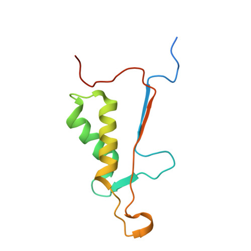
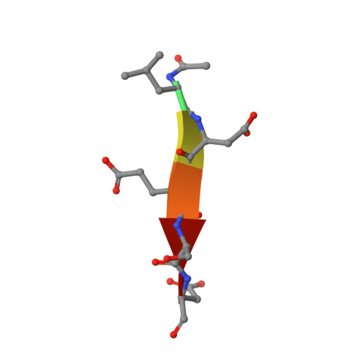
Structural basis for executioner caspase recognition of P5 position in substrates.

Fu, G.Chumanevich, A.A.Agniswamy, J.Fang, B.Harrison, R.W.Weber, I.T.

(2008) Apoptosis 13: 1291-1302

  • DOI: https://doi.org/10.1007/s10495-008-0259-9
  • Primary Citation of Related Structures:  
    3EDQ, 3EDR

  • PubMed Abstract: 

    Caspase-3, -6 and -7 cleave many proteins at specific sites to induce apoptosis. Their recognition of the P5 position in substrates has been investigated by kinetics, modeling and crystallography. Caspase-3 and -6 recognize P5 in pentapeptides as shown by enzyme activity data and interactions observed in the crystal structure of caspase-3/LDESD and in a model for caspase-6. In caspase-3 the P5 main-chain was anchored by interactions with Ser209 in loop-3 and the P5 Leu side-chain interacted with Phe250 and Phe252 in loop-4 consistent with 50% increased hydrolysis of LDEVD relative to DEVD. Caspase-6 formed similar interactions and showed a preference for polar P5 in QDEVD likely due to interactions with polar Lys265 and hydrophobic Phe263 in loop-4. Caspase-7 exhibited no preference for P5 residue in agreement with the absence of P5 interactions in the caspase-7/LDESD crystal structure. Initiator caspase-8, with Pro in the P5-anchoring position and no loop-4, had only 20% activity on tested pentapeptides relative to DEVD. Therefore, caspases-3 and -6 bind P5 using critical loop-3 anchoring Ser/Thr and loop-4 side-chain interactions, while caspase-7 and -8 lack P5-binding residues.


  • Organizational Affiliation

    Department of Biology, Molecular Basis of Disease Program, Georgia State University, Atlanta, GA 30303, USA.


Macromolecules
Find similar proteins by:  (by identity cutoff)  |  3D Structure
Entity ID: 1
MoleculeChains Sequence LengthOrganismDetailsImage
Caspase-3
A, C
147Homo sapiensMutation(s): 0 
Gene Names: CASP3CPP32
EC: 3.4.22.56
UniProt & NIH Common Fund Data Resources
Find proteins for P42574 (Homo sapiens)
Explore P42574 
Go to UniProtKB:  P42574
PHAROS:  P42574
GTEx:  ENSG00000164305 
Entity Groups  
Sequence Clusters30% Identity50% Identity70% Identity90% Identity95% Identity100% Identity
UniProt GroupP42574
Sequence Annotations
Expand
  • Reference Sequence
Find similar proteins by:  (by identity cutoff)  |  3D Structure
Entity ID: 2
MoleculeChains Sequence LengthOrganismDetailsImage
Caspase-3
B, D
108Homo sapiensMutation(s): 0 
Gene Names: CASP3CPP32
EC: 3.4.22.56
UniProt & NIH Common Fund Data Resources
Find proteins for P42574 (Homo sapiens)
Explore P42574 
Go to UniProtKB:  P42574
PHAROS:  P42574
GTEx:  ENSG00000164305 
Entity Groups  
Sequence Clusters30% Identity50% Identity70% Identity90% Identity95% Identity100% Identity
UniProt GroupP42574
Sequence Annotations
Expand
  • Reference Sequence

Find similar proteins by:  Sequence   |   3D Structure  

Entity ID: 3
MoleculeChains Sequence LengthOrganismDetailsImage
AC-LDESD-CHO peptide
E, F
6N/AMutation(s): 0 
Sequence Annotations
Expand
  • Reference Sequence
Small Molecules
Modified Residues  1 Unique
IDChains TypeFormula2D DiagramParent
ASA
Query on ASA
E, F
L-PEPTIDE LINKINGC4 H7 N O3ASP
Experimental Data & Validation

Experimental Data

  • Method: X-RAY DIFFRACTION
  • Resolution: 1.61 Å
  • R-Value Free: 0.223 
  • R-Value Work: 0.177 
  • R-Value Observed: 0.212 
  • Space Group: P 21 21 21
Unit Cell:
Length ( Å )Angle ( ˚ )
a = 67.375α = 90
b = 93.562β = 90
c = 97.617γ = 90
Software Package:
Software NamePurpose
MAR345data collection
PHASERphasing
SHELXL-97refinement
HKL-2000data scaling

Structure Validation

View Full Validation Report


Entry History 

Deposition Data

  • Released Date: 2008-10-28 
  • Deposition Author(s): Fu, G.

Revision History  (Full details and data files)

  • Version 1.0: 2008-10-28
    Type: Initial release
  • Version 1.1: 2011-07-13
    Changes: Atomic model, Database references, Derived calculations, Non-polymer description, Structure summary, Version format compliance
  • Version 1.2: 2017-10-25
    Changes: Refinement description
  • Version 1.3: 2023-08-30
    Changes: Data collection, Database references, Derived calculations, Refinement description
  • Version 1.4: 2023-11-15
    Changes: Data collection