Crystal structures of phosphotransferase system enzymes PtxB (IIB(Asc)) and PtxA (IIA(Asc)) from Streptococcus mutans
Lei, J., Li, L.F., Su, X.D.(2009) J Mol Biol 386: 465-475
- PubMed: 19135450
- DOI: https://doi.org/10.1016/j.jmb.2008.12.046
- Primary Citation of Related Structures:
3BJV, 3CZC - PubMed Abstract:
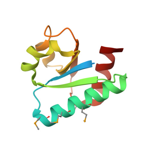
Streptococcus mutans is the primary etiological agent of dental caries in man and other mammalian organisms. This bacterium metabolizes carbohydrates actively and thrives under anaerobic conditions by fermenting l-ascorbate (Asc) via the sga operon, which includes SgaT, PtxB, and PtxA. These three proteins are members of the Asc family of enzyme II (EII) complexes of the bacterial phosphotransferase system. Here, we report the crystal structure of PtxB, solved by single-wavelength anomalous dispersion phasing, and that of PtxA, solved by molecular replacement, from S. mutans. PtxB provides the first crystal structure of an EIIB from the Asc family, composed of a central beta sheet of parallel strands flanked by alpha helices on both sides. The structure of PtxB is similar to the structures of IIB(Mtl) (IIB subunit of mannitol PTS) and IIB(Cel) (IIB subunit of cellobiose) in Escherichia coli despite the low sequence identity. PtxA adopts a globular alpha/beta sandwich structure. The phosphorylation-site His68 is situated between beta2 and beta3, within a hydrophobic pocket. We found that the hydrogen bond on N(delta1) of the active-site histidine is a common means of ensuring that phosphate is on the correct N(varepsilon2) site in many EIIA families. Finally, a model of the PtxB-PtxA complex was constructed, and a PtxA-phospho-PtxB state is proposed. Analyses of the two structures shed light on the catalytic mechanism of the phosphotransferase system.
Organizational Affiliation:
Peking University, Beijing, People's Republic of China.