3AWE

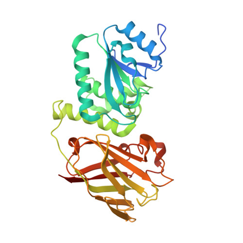
Crystal structure of Pten-like domain of Ci-VSP (248-576)


Experimental Data Snapshot

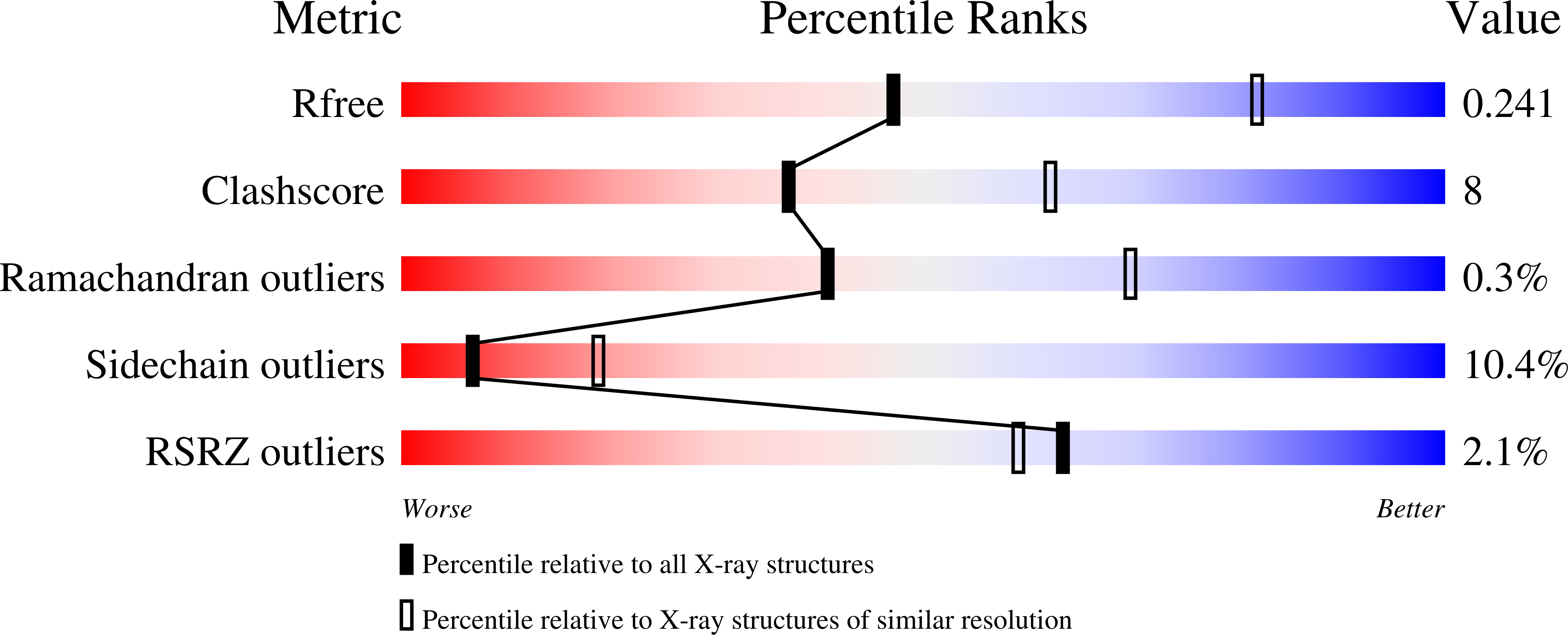
  • Method: X-RAY DIFFRACTION
  • Resolution: 2.77 Å
  • R-Value Free: 0.253 
  • R-Value Work: 0.208 
  • R-Value Observed: 0.210 

wwPDB Validation   3D Report Full Report


This is version 1.4 of the entry. See complete history


Literature

Crystal structure of the cytoplasmic phosphatase and tensin homolog (PTEN)-like region of Ciona intestinalis voltage-sensing phosphatase provides insight into substrate specificity and redox regulation of the phosphoinositide phosphatase activity

Matsuda, M.Takeshita, K.Kurokawa, T.Sakata, S.Suzuki, M.Yamashita, E.Okamura, Y.Nakagawa, A.

(2011) J Biol Chem 286: 23368-23377

  • DOI: https://doi.org/10.1074/jbc.M110.214361
  • Primary Citation of Related Structures:  
    3AWE, 3AWF, 3AWG

  • PubMed Abstract: 

    Ciona intestinalis voltage-sensing phosphatase (Ci-VSP) has a transmembrane voltage sensor domain and a cytoplasmic region sharing similarity to the phosphatase and tensin homolog (PTEN). It dephosphorylates phosphatidylinositol 4,5-bisphosphate and phosphatidylinositol 3,4,5-trisphosphate upon membrane depolarization. The cytoplasmic region is composed of a phosphatase domain and a putative membrane interaction domain, C2. Here we determined the crystal structures of the Ci-VSP cytoplasmic region in three distinct constructs, wild-type (248-576), wild-type (236-576), and G365A mutant (248-576). The crystal structure of WT-236 and G365A-248 had the disulfide bond between the catalytic residue Cys-363 and the adjacent residue Cys-310. On the other hand, the disulfide bond was not present in the crystal structure of WT-248. These suggest the possibility that Ci-VSP is regulated by reactive oxygen species as found in PTEN. These structures also revealed that the conformation of the TI loop in the active site of the Ci-VSP cytoplasmic region was distinct from the corresponding region of PTEN; Ci-VSP has glutamic acid (Glu-411) in the TI loop, orienting toward the center of active site pocket. Mutation of Glu-411 led to acquirement of increased activity toward phosphatidylinositol 3,5-bisphosphate, suggesting that this site is required for determining substrate specificity. Our results provide the basic information of the enzymatic mechanism of Ci-VSP.


  • Organizational Affiliation

    Institute for Protein Research, Graduate School of Medicine, Osaka University, Suita, Osaka 565-0871, Japan.


Macromolecules
Find similar proteins by:  (by identity cutoff)  |  3D Structure
Entity ID: 1
MoleculeChains Sequence LengthOrganismDetailsImage
Voltage-sensor containing phosphatase
A, B, C
334Ciona intestinalisMutation(s): 0 
Gene Names: Ci-VSP
EC: 3.1.3
UniProt
Find proteins for Q4W8A1 (Ciona intestinalis)
Explore Q4W8A1 
Go to UniProtKB:  Q4W8A1
Entity Groups  
Sequence Clusters30% Identity50% Identity70% Identity90% Identity95% Identity100% Identity
UniProt GroupQ4W8A1
Sequence Annotations
Expand
  • Reference Sequence
Small Molecules
Ligands 3 Unique
IDChains Name / Formula / InChI Key2D Diagram3D Interactions
SO4
Query on SO4

Download Ideal Coordinates CCD File 
D [auth A]
E [auth A]
G [auth B]
H [auth B]
J [auth C]
D [auth A],
E [auth A],
G [auth B],
H [auth B],
J [auth C],
K [auth C],
L [auth C]
SULFATE ION
O4 S
QAOWNCQODCNURD-UHFFFAOYSA-L
ACY
Query on ACY

Download Ideal Coordinates CCD File 
I [auth C]ACETIC ACID
C2 H4 O2
QTBSBXVTEAMEQO-UHFFFAOYSA-N
NA
Query on NA

Download Ideal Coordinates CCD File 
F [auth A]SODIUM ION
Na
FKNQFGJONOIPTF-UHFFFAOYSA-N
Experimental Data & Validation

Experimental Data

  • Method: X-RAY DIFFRACTION
  • Resolution: 2.77 Å
  • R-Value Free: 0.253 
  • R-Value Work: 0.208 
  • R-Value Observed: 0.210 
  • Space Group: P 21 21 2
Unit Cell:
Length ( Å )Angle ( ˚ )
a = 131.207α = 90
b = 176.527β = 90
c = 50.794γ = 90
Software Package:
Software NamePurpose
BALBESphasing
REFMACrefinement
DENZOdata reduction
SCALEPACKdata scaling

Structure Validation

View Full Validation Report


Entry History 

Deposition Data

Revision History  (Full details and data files)

  • Version 1.0: 2011-05-04
    Type: Initial release
  • Version 1.1: 2011-07-13
    Changes: Version format compliance
  • Version 1.2: 2011-12-21
    Changes: Database references
  • Version 1.3: 2017-10-11
    Changes: Refinement description
  • Version 1.4: 2023-11-01
    Changes: Data collection, Database references, Derived calculations, Refinement description