3VVW

NDP52 in complex with LC3C


Experimental Data Snapshot

  • Method: X-RAY DIFFRACTION
  • Resolution: 2.50 Å
  • R-Value Free: 0.252 
  • R-Value Work: 0.210 
  • R-Value Observed: 0.212 

wwPDB Validation   3D Report Full Report


This is version 1.1 of the entry. See complete history


Literature

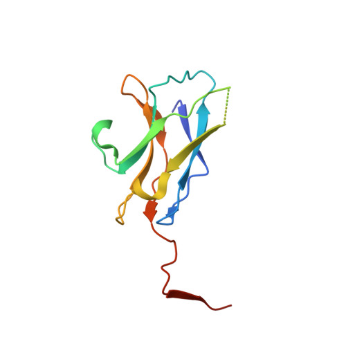
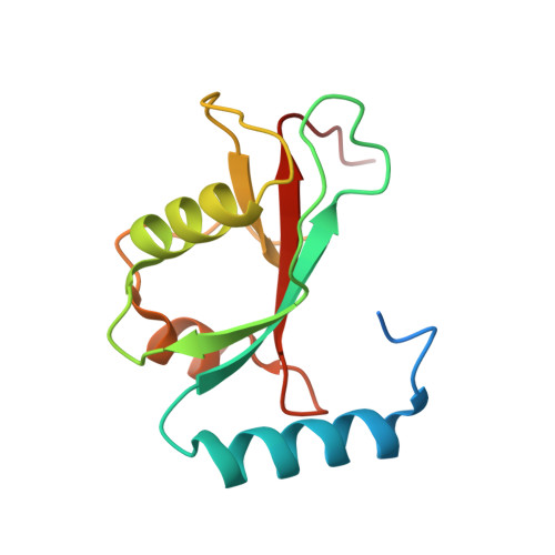
LC3C, bound selectively by a noncanonical LIR motif in NDP52, is required for antibacterial autophagy

Muhlinen, N.V.Akutsu, M.Ravenhill, B.J.Foeglein, A.Bloor, S.Rutherford, T.J.Freund, S.M.Komander, D.Randow, F.

(2012) Mol Cell 48: 329-342

  • DOI: https://doi.org/10.1016/j.molcel.2012.08.024
  • Primary Citation of Related Structures:  
    3VVV, 3VVW

  • PubMed Abstract: 

    Autophagy protects cellular homeostasis by capturing cytosolic components and invading pathogens for lysosomal degradation. Autophagy receptors target cargo to autophagy by binding ATG8 on autophagosomal membranes. The expansion of the ATG8 family in higher eukaryotes suggests that specific interactions with autophagy receptors facilitate differential cargo handling. However, selective interactors of ATG8 orthologs are unknown. Here we show that the selectivity of the autophagy receptor NDP52 for LC3C is crucial for innate immunity since cells lacking either protein cannot protect their cytoplasm against Salmonella. LC3C is required for antibacterial autophagy because in its absence the remaining ATG8 orthologs do not support efficient antibacterial autophagy. Structural analysis revealed that the selectivity of NDP52 for LC3C is conferred by a noncanonical LIR, in which lack of an aromatic residue is balanced by LC3C-specific interactions. Our report illustrates that specificity in the interaction between autophagy receptors and autophagy machinery is of functional importance to execute selective autophagy.


  • Organizational Affiliation

    MRC Laboratory of Molecular Biology, Division of Protein and Nucleic Acid Chemistry, Cambridge CB2 0QH, UK.


Macromolecules
Find similar proteins by:  (by identity cutoff)  |  3D Structure
Entity ID: 1
MoleculeChains Sequence LengthOrganismDetailsImage
Calcium-binding and coiled-coil domain-containing protein 2123Homo sapiensMutation(s): 0 
Gene Names: CALCOCO2NDP52
UniProt & NIH Common Fund Data Resources
Find proteins for Q13137 (Homo sapiens)
Explore Q13137 
Go to UniProtKB:  Q13137
PHAROS:  Q13137
GTEx:  ENSG00000136436 
Entity Groups  
Sequence Clusters30% Identity50% Identity70% Identity90% Identity95% Identity100% Identity
UniProt GroupQ13137
Sequence Annotations
Expand
  • Reference Sequence
Find similar proteins by:  (by identity cutoff)  |  3D Structure
Entity ID: 2
MoleculeChains Sequence LengthOrganismDetailsImage
Microtubule-associated proteins 1A/1B light chain 3C128Homo sapiensMutation(s): 0 
Gene Names: MAP1LC3C
UniProt & NIH Common Fund Data Resources
Find proteins for Q9BXW4 (Homo sapiens)
Explore Q9BXW4 
Go to UniProtKB:  Q9BXW4
PHAROS:  Q9BXW4
GTEx:  ENSG00000197769 
Entity Groups  
Sequence Clusters30% Identity50% Identity70% Identity90% Identity95% Identity100% Identity
UniProt GroupQ9BXW4
Sequence Annotations
Expand
  • Reference Sequence
Experimental Data & Validation

Experimental Data

  • Method: X-RAY DIFFRACTION
  • Resolution: 2.50 Å
  • R-Value Free: 0.252 
  • R-Value Work: 0.210 
  • R-Value Observed: 0.212 
  • Space Group: C 1 2 1
Unit Cell:
Length ( Å )Angle ( ˚ )
a = 195.72α = 90
b = 37.42β = 94.66
c = 40.28γ = 90
Software Package:
Software NamePurpose
PHENIXrefinement
PDB_EXTRACTdata extraction
ADSCdata collection
MOSFLMdata reduction
SCALAdata scaling
PHENIXphasing

Structure Validation

View Full Validation Report


Entry History 

Deposition Data

Revision History  (Full details and data files)

  • Version 1.0: 2013-02-27
    Type: Initial release
  • Version 1.1: 2018-06-13
    Changes: Data collection, Derived calculations