


Crystal Structure of a Parallel Coiled-Coil Dimerization Domain from the Voltage-Gated Proton Channel (Mutation/C245S)
Fujiwara, Y., Takeshita, K., Kobayashi, M., Okamura, Y., Nakagawa, A.To be published.
Experimental Data Snapshot
wwPDB Validation 3D Report Full Report

Entity ID: 1 | |||||
|---|---|---|---|---|---|
| Molecule | Chains | Sequence Length | Organism | Details | Image |
| Voltage-gated hydrogen channel 1 | A, B [auth C], C [auth B], D | 51 | Mus musculus | Mutation(s): 1 Gene Names: Bts, Hvcn1, Vsop | 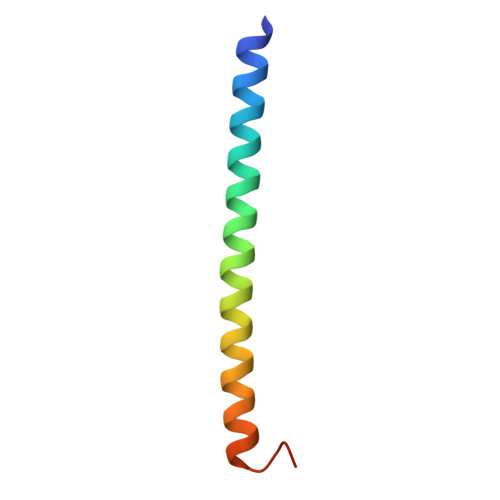 |
UniProt | |||||
Find proteins for Q3U2S8 (Mus musculus) Explore Q3U2S8 Go to UniProtKB: Q3U2S8 | |||||
Entity Groups | |||||
| Sequence Clusters | 30% Identity50% Identity70% Identity90% Identity95% Identity100% Identity | ||||
| UniProt Group | Q3U2S8 | ||||
Sequence AnnotationsExpand | |||||
| |||||
| Length ( Å ) | Angle ( ˚ ) |
|---|---|
| a = 40.126 | α = 90 |
| b = 53.868 | β = 90 |
| c = 81.413 | γ = 90 |
| Software Name | Purpose |
|---|---|
| REFMAC | refinement |
| DENZO | data reduction |
| SCALEPACK | data scaling |
| REFMAC | phasing |
RCSB PDB (citation) is hosted by
RCSB PDB is a member of the

