The 1.35 A resolution structure of the phosphatase domain of the suppressor of T-cell receptor signaling protein in complex with sulfate.
Jakoncic, J., Sondgeroth, B., Carpino, N., Nassar, N.(2010) Acta Crystallogr Sect F Struct Biol Cryst Commun 66: 643-647
- PubMed: 20516590
- DOI: https://doi.org/10.1107/S1744309110014259
- Primary Citation of Related Structures:
3MBK - PubMed Abstract:
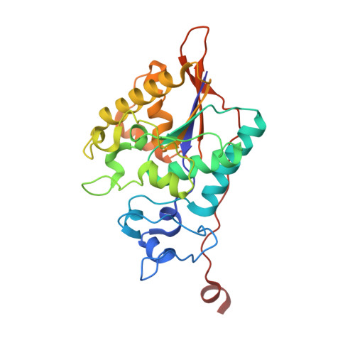
The suppressor of T-cell signaling (Sts) proteins are multidomain proteins that negatively regulate the signaling of membrane-bound receptors, including the T-cell receptor (TCR) and the epidermal growth-factor receptor (EGFR). They contain at their C-terminus a 2H-phosphatase homology (PGM) domain that is responsible for their protein tyrosine phosphatase activity. Here, the crystal structure of the phosphatase domain of Sts-1, Sts-1(PGM), was determined at pH 4.6. The asymmetric unit contains two independent molecules and each active site is occupied by a sulfate ion. Each sulfate is located at the phosphate-binding site and makes similar interactions with the catalytic residues. The structure suggests an explanation for the lower Michaelis-Menten constants at acidic pH.
Organizational Affiliation:
Brookhaven National Laboratory, National Synchrotron Light Source, Building 725, Upton, NY 11973, USA.