3IM6

Crystal structure of mouse Ryanodine Receptor 2 mutant V186M


Experimental Data Snapshot

  • Method: X-RAY DIFFRACTION
  • Resolution: 1.70 Å
  • R-Value Free: 0.244 
  • R-Value Work: 0.201 
  • R-Value Observed: 0.203 

wwPDB Validation   3D Report Full Report


This is version 1.3 of the entry. See complete history


Literature

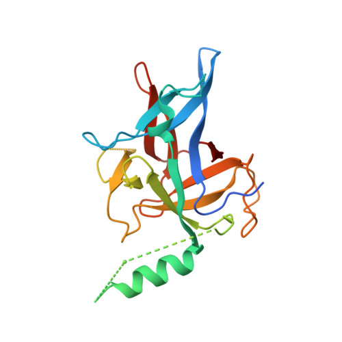
Crystal structures of the N-terminal domains of cardiac and skeletal muscle ryanodine receptors: insights into disease mutations.

Lobo, P.A.Van Petegem, F.

(2009) Structure 17: 1505-1514

  • DOI: https://doi.org/10.1016/j.str.2009.08.016
  • Primary Citation of Related Structures:  
    3ILA, 3IM5, 3IM6, 3IM7

  • PubMed Abstract: 

    Ryanodine receptors (RyRs) are channels governing the release of Ca(2+) from the sarcoplasmic or endoplasmic reticulum. They are required for the contraction of both skeletal (RyR1) and cardiac (RyR2) muscles. Mutations in both RyR1 and RyR2 have been associated with severe genetic disorders, but high-resolution data describing the disease variants in detail have been lacking. Here we present the crystal structures of the N-terminal domains of both RyR2 (1-217) and RyR1 (9-205) at 2.55 A and 2.9 A, respectively. The domains map in a hot spot region for disease mutations. Both structures consist of a core beta trefoil domain flanked by an alpha helix. Crystal structures of two RyR2 disease mutants, A77V (2.2 A) and V186M (1.7 A), show that the mutations cause distinct local changes in the surface of the protein. A RyR2 deletion mutant causes significant changes in the thermal stability. The disease positions highlight two putative binding interfaces required for normal RyR function.


  • Organizational Affiliation

    University of British Columbia, Department of Biochemistry and Molecular Biology, Vancouver, BC V6T 1Z3 Canada.


Macromolecules
Find similar proteins by:  (by identity cutoff)  |  3D Structure
Entity ID: 1
MoleculeChains Sequence LengthOrganismDetailsImage
Cardiac Ca2+ release channel217Mus musculusMutation(s): 1 
Gene Names: Ryr2
UniProt & NIH Common Fund Data Resources
Find proteins for E9Q401 (Mus musculus)
Explore E9Q401 
Go to UniProtKB:  E9Q401
IMPC:  MGI:99685
Entity Groups  
Sequence Clusters30% Identity50% Identity70% Identity90% Identity95% Identity100% Identity
UniProt GroupE9Q401
Sequence Annotations
Expand
  • Reference Sequence
Small Molecules
Ligands 1 Unique
IDChains Name / Formula / InChI Key2D Diagram3D Interactions
SO4
Query on SO4

Download Ideal Coordinates CCD File 
B [auth A]SULFATE ION
O4 S
QAOWNCQODCNURD-UHFFFAOYSA-L
Experimental Data & Validation

Experimental Data

  • Method: X-RAY DIFFRACTION
  • Resolution: 1.70 Å
  • R-Value Free: 0.244 
  • R-Value Work: 0.201 
  • R-Value Observed: 0.203 
  • Space Group: I 41
Unit Cell:
Length ( Å )Angle ( ˚ )
a = 112.512α = 90
b = 112.512β = 90
c = 36.751γ = 90
Software Package:
Software NamePurpose
HKL-2000data collection
PHASERphasing
REFMACrefinement
HKL-2000data reduction
HKL-2000data scaling

Structure Validation

View Full Validation Report


Entry History 

Deposition Data

Revision History  (Full details and data files)

  • Version 1.0: 2009-11-17
    Type: Initial release
  • Version 1.1: 2011-07-13
    Changes: Version format compliance
  • Version 1.2: 2021-10-13
    Changes: Database references, Derived calculations
  • Version 1.3: 2023-09-06
    Changes: Data collection, Refinement description