Molecular basis for insulin fibril assembly.
Ivanova, M.I., Sievers, S.A., Sawaya, M.R., Wall, J.S., Eisenberg, D.(2009) Proc Natl Acad Sci U S A 106: 18990-18995
- PubMed: 19864624
- DOI: https://doi.org/10.1073/pnas.0910080106
- Primary Citation of Related Structures:
3HYD - PubMed Abstract:
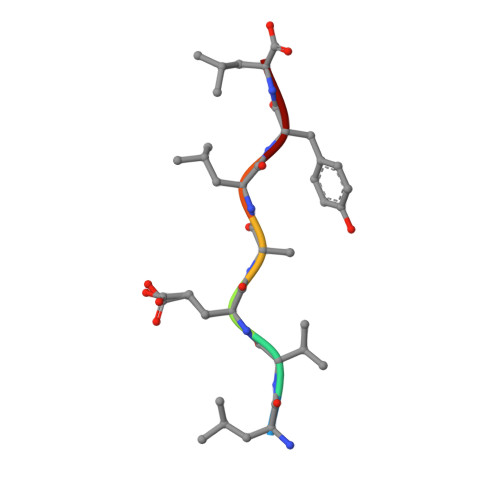
In the rare medical condition termed injection amyloidosis, extracellular fibrils of insulin are observed. We found that the segment of the insulin B-chain with sequence LVEALYL is the smallest segment that both nucleates and inhibits the fibrillation of full-length insulin in a molar ratio-dependent manner, suggesting that this segment is central to the cross-beta spine of the insulin fibril. In isolation from the rest of the protein, LVEALYL forms microcrystalline aggregates with fibrillar morphology, the structure of which we determined to 1 A resolution. The LVEALYL segments are stacked into pairs of tightly interdigitated beta-sheets, each pair displaying the dry steric zipper interface typical of amyloid-like fibrils. This structure leads to a model for fibrils of human insulin consistent with electron microscopic, x-ray fiber diffraction, and biochemical studies.
Organizational Affiliation:
Howard Hughes Medical Institute, UCLA-DOE Institute for Genomics and Proteomics, Los Angeles CA 90095-1570, USA.