2YII

Manipulating the regioselectivity of phenylalanine aminomutase: new insights into the reaction mechanism of MIO-dependent enzymes from structure-guided directed evolution


Experimental Data Snapshot

  • Method: X-RAY DIFFRACTION
  • Resolution: 2.18 Å
  • R-Value Free: 0.214 
  • R-Value Work: 0.179 
  • R-Value Observed: 0.181 

wwPDB Validation   3D Report Full Report


This is version 2.1 of the entry. See complete history


Literature

Mechanism-Inspired Engineering of Phenylalanine Aminomutase for Enhanced Beta-Regioselective Asymmetric Amination of Cinnamates.

Wu, B.Szymanski, W.Wybenga, G.G.Heberling, M.M.Bartsch, S.De Wildeman, S.Poelarends, G.J.Feringa, B.L.Dijkstra, B.W.Janssen, D.B.

(2012) Angew Chem Int Ed Engl 51: 482

  • DOI: https://doi.org/10.1002/anie.201106372
  • Primary Citation of Related Structures:  
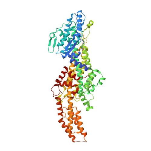
    2YII

  • PubMed Abstract: 

    Turn to switch: A mutant of phenylalanine aminomutase was engineered that can catalyze the regioselective amination of cinnamate derivatives (see scheme, red) to, for example, β-amino acids. This regioselectivity, along with the X-ray crystal structures, suggests two distinct carboxylate binding modes differentiated by C(β)-C(ipso) bond rotation, which determines if β- (see scheme) or α-addition takes place.


  • Organizational Affiliation

    Department of Biochemistry, Groningen Biomolecular Sciences and Biotechnology Institute, University of Groningen, Nijenborgh 4, 9747 AG, Groningen, The Netherlands.


Macromolecules
Find similar proteins by:  (by identity cutoff)  |  3D Structure
Entity ID: 1
MoleculeChains Sequence LengthOrganismDetailsImage
PHENYLALANINE AMMONIA-LYASE
A, B, C, D
705Taxus chinensisMutation(s): 1 
EC: 5.4.3 (PDB Primary Data), 4.3.1.24 (PDB Primary Data)
UniProt
Find proteins for Q68G84 (Taxus chinensis)
Explore Q68G84 
Go to UniProtKB:  Q68G84
Entity Groups  
Sequence Clusters30% Identity50% Identity70% Identity90% Identity95% Identity100% Identity
UniProt GroupQ68G84
Sequence Annotations
Expand
  • Reference Sequence
Small Molecules
Ligands 3 Unique
IDChains Name / Formula / InChI Key2D Diagram3D Interactions
GOL
Query on GOL

Download Ideal Coordinates CCD File 
J [auth B]GLYCEROL
C3 H8 O3
PEDCQBHIVMGVHV-UHFFFAOYSA-N
BME
Query on BME

Download Ideal Coordinates CCD File 
E [auth A]
F [auth A]
H [auth B]
I [auth B]
L [auth C]
E [auth A],
F [auth A],
H [auth B],
I [auth B],
L [auth C],
M [auth C],
O [auth D],
P [auth D]
BETA-MERCAPTOETHANOL
C2 H6 O S
DGVVWUTYPXICAM-UHFFFAOYSA-N
FMT
Query on FMT

Download Ideal Coordinates CCD File 
G [auth A],
K [auth B],
N [auth C],
Q [auth D]
FORMIC ACID
C H2 O2
BDAGIHXWWSANSR-UHFFFAOYSA-N
Modified Residues  1 Unique
IDChains TypeFormula2D DiagramParent
MDO
Query on MDO
A, B, C, D
L-PEPTIDE LINKINGC8 H11 N3 O3ALA, SER, GLY
Experimental Data & Validation

Experimental Data

  • Method: X-RAY DIFFRACTION
  • Resolution: 2.18 Å
  • R-Value Free: 0.214 
  • R-Value Work: 0.179 
  • R-Value Observed: 0.181 
  • Space Group: P 1 21 1
Unit Cell:
Length ( Å )Angle ( ˚ )
a = 99.454α = 90
b = 145.999β = 99.51
c = 99.68γ = 90
Software Package:
Software NamePurpose
REFMACrefinement
XDSdata reduction
SCALAdata scaling
PHASERphasing

Structure Validation

View Full Validation Report


Entry History 

Deposition Data

Revision History  (Full details and data files)

  • Version 1.0: 2011-11-09
    Type: Initial release
  • Version 1.1: 2012-01-25
    Changes: Other
  • Version 1.2: 2013-01-30
    Changes: Database references
  • Version 1.3: 2019-10-23
    Changes: Data collection, Database references, Derived calculations, Other
  • Version 2.0: 2023-11-15
    Changes: Atomic model, Data collection, Database references, Derived calculations
  • Version 2.1: 2023-12-20
    Changes: Refinement description