2WBW

Ad37 fibre head in complex with CAR D1 and sialic acid


Experimental Data Snapshot

  • Method: X-RAY DIFFRACTION
  • Resolution: 1.55 Å
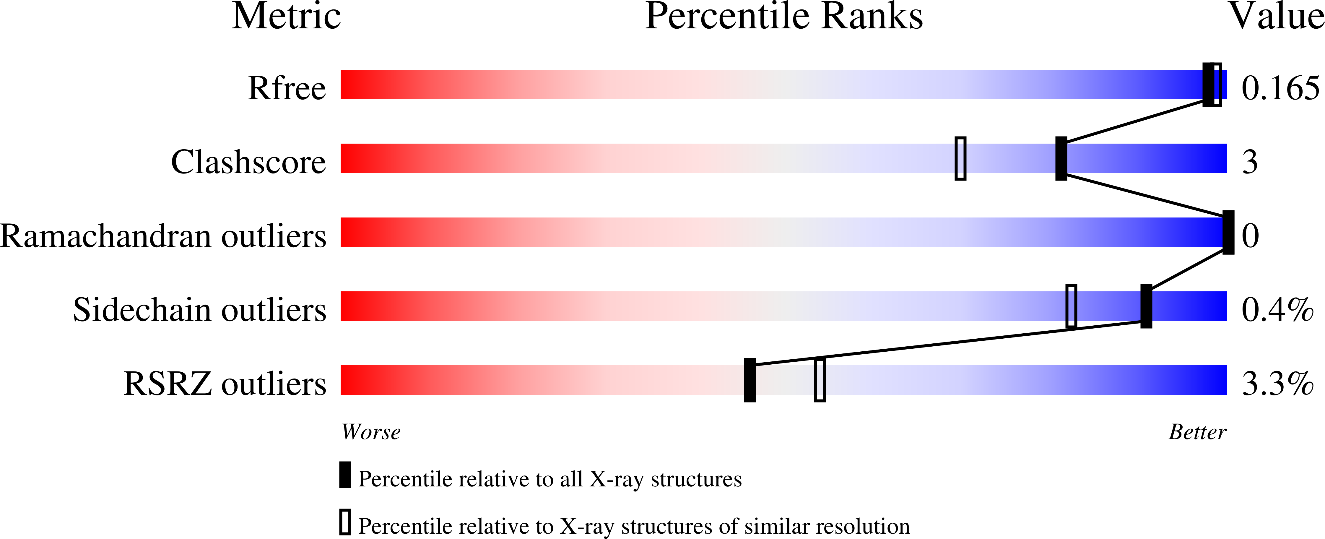
  • R-Value Free: 0.176 
  • R-Value Work: 0.171 
  • R-Value Observed: 0.171 

wwPDB Validation   3D Report Full Report


Ligand Structure Quality Assessment 


This is version 1.3 of the entry. See complete history


Literature

The Cell Adhesion Molecule "Car" and Sialic Acid on Human Erythrocytes Influence Adenovirus in Vivo Biodistribution.

Seiradake, E.Henaff, D.Wodrich, H.Billet, O.Perreau, M.Hippert, C.Mennechet, F.Schoehn, G.Lortat-Jacob, H.Dreja, H.Ibanes, S.Kalatzis, V.Wang, J.P.Finberg, R.W.Cusack, S.Kremer, E.J.

(2009) PLoS Pathog 5: 00277

  • DOI: https://doi.org/10.1371/journal.ppat.1000277
  • Primary Citation of Related Structures:  
    2W9L, 2WBV, 2WBW

  • PubMed Abstract: 

    Although it has been known for 50 years that adenoviruses (Ads) interact with erythrocytes ex vivo, the molecular and structural basis for this interaction, which has been serendipitously exploited for diagnostic tests, is unknown. In this study, we characterized the interaction between erythrocytes and unrelated Ad serotypes, human 5 (HAd5) and 37 (HAd37), and canine 2 (CAV-2). While these serotypes agglutinate human erythrocytes, they use different receptors, have different tropisms and/or infect different species. Using molecular, biochemical, structural and transgenic animal-based analyses, we found that the primary erythrocyte interaction domain for HAd37 is its sialic acid binding site, while CAV-2 binding depends on at least three factors: electrostatic interactions, sialic acid binding and, unexpectedly, binding to the coxsackievirus and adenovirus receptor (CAR) on human erythrocytes. We show that the presence of CAR on erythrocytes leads to prolonged in vivo blood half-life and significantly reduced liver infection when a CAR-tropic Ad is injected intravenously. This study provides i) a molecular and structural rationale for Ad-erythrocyte interactions, ii) a basis to improve vector-mediated gene transfer and iii) a mechanism that may explain the biodistribution and pathogenic inconsistencies found between human and animal models.


  • Organizational Affiliation

    European Molecular Biology Laboratory, Grenoble Outstation, Grenoble, France.


Macromolecules
Find similar proteins by:  (by identity cutoff)  |  3D Structure
Entity ID: 1
MoleculeChains Sequence LengthOrganismDetailsImage
FIBER PROTEIN194Human adenovirus D37Mutation(s): 0 
UniProt
Find proteins for Q64823 (Human adenovirus D37)
Explore Q64823 
Go to UniProtKB:  Q64823
Entity Groups  
Sequence Clusters30% Identity50% Identity70% Identity90% Identity95% Identity100% Identity
UniProt GroupQ64823
Sequence Annotations
Expand
  • Reference Sequence
Find similar proteins by:  (by identity cutoff)  |  3D Structure
Entity ID: 2
MoleculeChains Sequence LengthOrganismDetailsImage
COXSACKIEVIRUS AND ADENOVIRUS RECEPTOR128Homo sapiensMutation(s): 0 
UniProt & NIH Common Fund Data Resources
Find proteins for P78310 (Homo sapiens)
Explore P78310 
Go to UniProtKB:  P78310
PHAROS:  P78310
GTEx:  ENSG00000154639 
Entity Groups  
Sequence Clusters30% Identity50% Identity70% Identity90% Identity95% Identity100% Identity
UniProt GroupP78310
Sequence Annotations
Expand
  • Reference Sequence
Small Molecules
Ligands 2 Unique
IDChains Name / Formula / InChI Key2D Diagram3D Interactions
SIA
Query on SIA

Download Ideal Coordinates CCD File 
C [auth A]N-acetyl-alpha-neuraminic acid
C11 H19 N O9
SQVRNKJHWKZAKO-YRMXFSIDSA-N
CA
Query on CA

Download Ideal Coordinates CCD File 
D [auth B]CALCIUM ION
Ca
BHPQYMZQTOCNFJ-UHFFFAOYSA-N
Experimental Data & Validation

Experimental Data

  • Method: X-RAY DIFFRACTION
  • Resolution: 1.55 Å
  • R-Value Free: 0.176 
  • R-Value Work: 0.171 
  • R-Value Observed: 0.171 
  • Space Group: I 2 3
Unit Cell:
Length ( Å )Angle ( ˚ )
a = 132.01α = 90
b = 132.01β = 90
c = 132.01γ = 90
Software Package:
Software NamePurpose
REFMACrefinement
XDSdata reduction
XSCALEdata scaling
PHASERphasing

Structure Validation

View Full Validation Report


Ligand Structure Quality Assessment 


Entry History 

Revision History  (Full details and data files)

  • Version 1.0: 2009-03-17
    Type: Initial release
  • Version 1.1: 2011-12-28
    Changes: Database references, Derived calculations, Non-polymer description, Other, Version format compliance
  • Version 1.2: 2020-07-29
    Type: Remediation
    Reason: Carbohydrate remediation
    Changes: Data collection, Derived calculations, Other, Structure summary
  • Version 1.3: 2023-12-13
    Changes: Data collection, Database references, Refinement description, Structure summary