2WAB

Structure of an active site mutant of a family two carbohydrate esterase from Clostridium thermocellum in complex with celluohexase


Experimental Data Snapshot

  • Method: X-RAY DIFFRACTION
  • Resolution: 1.90 Å
  • R-Value Free: 0.203 
  • R-Value Work: 0.156 
  • R-Value Observed: 0.158 

wwPDB Validation   3D Report Full Report


This is version 2.0 of the entry. See complete history


Literature

The Active Site of a Carbohydrate Esterase Displays Divergent Catalytic and Noncatalytic Binding Functions.

Montanier, C.Money, V.A.Pires, V.M.R.Flint, J.E.Pinheiro, B.A.Goyal, A.Prates, J.A.M.Izumi, A.Stalbrand, H.Morland, C.Cartmell, A.Kolenova, K.Topakas, E.Dodson, E.J.Bolam, D.N.Davies, G.J.Fontes, C.M.G.A.Gilbert, H.J.

(2009) PLoS Biol 7: E71

  • DOI: https://doi.org/10.1371/journal.pbio.1000071
  • Primary Citation of Related Structures:  
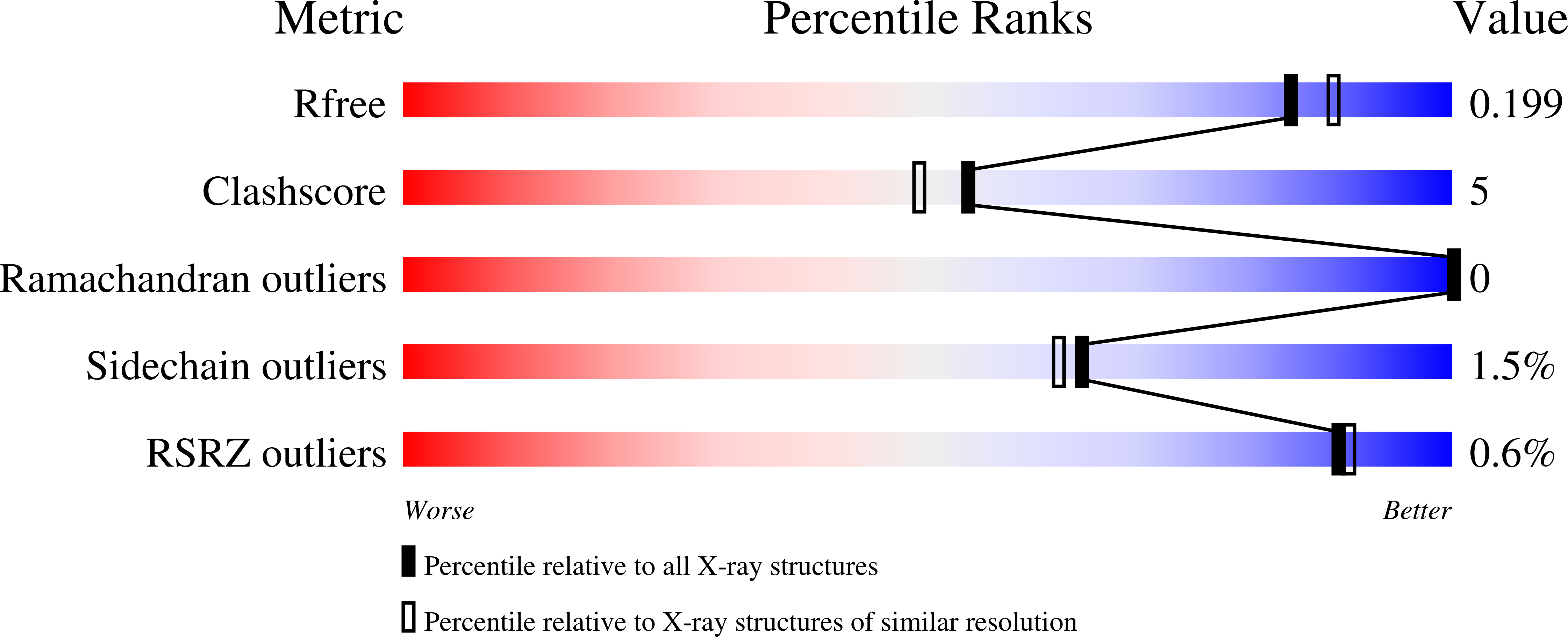
    2W9X, 2WAA, 2WAB, 2WAO

  • PubMed Abstract: 

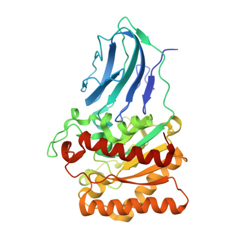
    Multifunctional proteins, which play a critical role in many biological processes, have typically evolved through the recruitment of different domains that have the required functional diversity. Thus the different activities displayed by these proteins are mediated by spatially distinct domains, consistent with the specific chemical requirements of each activity. Indeed, current evolutionary theory argues that the colocalization of diverse activities within an enzyme is likely to be a rare event, because it would compromise the existing activity of the protein. In contrast to this view, a potential example of multifunctional recruitment into a single protein domain is provided by CtCel5C-CE2, which contains an N-terminal module that displays cellulase activity and a C-terminal module, CtCE2, which exhibits a noncatalytic cellulose-binding function but also shares sequence identity with the CE2 family of esterases. Here we show that, unlike other CE2 members, the CtCE2 domain displays divergent catalytic esterase and noncatalytic carbohydrate binding functions. Intriguingly, these diverse activities are housed within the same site on the protein. Thus, a critical component of the active site of CtCE2, the catalytic Ser-His dyad, in harness with inserted aromatic residues, confers noncatalytic binding to cellulose whilst the active site of the domain retains its esterase activity. CtCE2 catalyses deacetylation of noncellulosic plant structural polysaccharides to deprotect these substrates for attack by other enzymes. Yet it also acts as a cellulose-binding domain, which promotes the activity of the appended cellulase on recalcitrant substrates. The CE2 family encapsulates the requirement for multiple activities by biocatalysts that attack challenging macromolecular substrates, including the grafting of a second, powerful and discrete noncatalytic binding functionality into the active site of an enzyme. This article provides a rare example of "gene sharing," where the introduction of a second functionality into the active site of an enzyme does not compromise the original activity of the biocatalyst.


  • Organizational Affiliation

    Institute for Cell and Molecular Biosciences, Newcastle University, The Medical School, Newcastle upon Tyne, UK.


Macromolecules
Find similar proteins by:  (by identity cutoff)  |  3D Structure
Entity ID: 1
MoleculeChains Sequence LengthOrganismDetailsImage
ENDOGLUCANASE E341Acetivibrio thermocellusMutation(s): 1 
EC: 3.2.1.4
UniProt
Find proteins for P10477 (Acetivibrio thermocellus (strain ATCC 27405 / DSM 1237 / JCM 9322 / NBRC 103400 / NCIMB 10682 / NRRL B-4536 / VPI 7372))
Explore P10477 
Go to UniProtKB:  P10477
Entity Groups  
Sequence Clusters30% Identity50% Identity70% Identity90% Identity95% Identity100% Identity
UniProt GroupP10477
Sequence Annotations
Expand
  • Reference Sequence
Oligosaccharides

Help

Entity ID: 2
MoleculeChains Length2D Diagram Glycosylation3D Interactions
beta-D-glucopyranose-(1-4)-beta-D-glucopyranose-(1-4)-beta-D-glucopyranose-(1-4)-beta-D-glucopyranose-(1-4)-beta-D-glucopyranose
B
5N/A
Glycosylation Resources
GlyTouCan:  G91683DU
GlyCosmos:  G91683DU
Small Molecules
Ligands 2 Unique
IDChains Name / Formula / InChI Key2D Diagram3D Interactions
IOD
Query on IOD

Download Ideal Coordinates CCD File 
I [auth A]
J [auth A]
K [auth A]
L [auth A]
M [auth A]
I [auth A],
J [auth A],
K [auth A],
L [auth A],
M [auth A],
N [auth A]
IODIDE ION
I
XMBWDFGMSWQBCA-UHFFFAOYSA-M
GOL
Query on GOL

Download Ideal Coordinates CCD File 
C [auth A]
D [auth A]
E [auth A]
F [auth A]
G [auth A]
C [auth A],
D [auth A],
E [auth A],
F [auth A],
G [auth A],
H [auth A]
GLYCEROL
C3 H8 O3
PEDCQBHIVMGVHV-UHFFFAOYSA-N
Biologically Interesting Molecules (External Reference) 1 Unique
Experimental Data & Validation

Experimental Data

  • Method: X-RAY DIFFRACTION
  • Resolution: 1.90 Å
  • R-Value Free: 0.203 
  • R-Value Work: 0.156 
  • R-Value Observed: 0.158 
  • Space Group: P 21 21 2
Unit Cell:
Length ( Å )Angle ( ˚ )
a = 41.158α = 90
b = 141.235β = 90
c = 58.161γ = 90
Software Package:
Software NamePurpose
REFMACrefinement
MOSFLMdata reduction
SCALAdata scaling
PHASERphasing

Structure Validation

View Full Validation Report


Entry History 

Revision History  (Full details and data files)

  • Version 1.0: 2009-03-24
    Type: Initial release
  • Version 1.1: 2011-05-08
    Changes: Version format compliance
  • Version 1.2: 2011-07-13
    Changes: Version format compliance
  • Version 1.3: 2018-05-02
    Changes: Data collection
  • Version 2.0: 2020-07-29
    Type: Remediation
    Reason: Carbohydrate remediation
    Changes: Advisory, Atomic model, Data collection, Derived calculations, Other, Structure summary