2W57

Crystal structure of the Vibrio cholerae ferric uptake regulator (Fur) reveals structural rearrangement of the DNA-binding domains


Experimental Data Snapshot

  • Method: X-RAY DIFFRACTION
  • Resolution: 2.60 Å
  • R-Value Free: 0.259 
  • R-Value Work: 0.216 
  • R-Value Observed: 0.218 

wwPDB Validation   3D Report Full Report


This is version 1.2 of the entry. See complete history


Literature

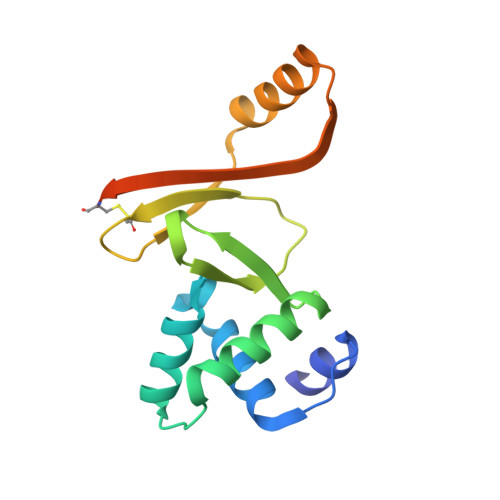
Crystal Structure of the Vibrio Cholerae Ferric Uptake Regulator (Fur) Reveals Insights Into Metal Co-Ordination.

Sheikh, M.A.Taylor, G.L.

(2009) Mol Microbiol 72: 1208

  • DOI: https://doi.org/10.1111/j.1365-2958.2009.06718.x
  • Primary Citation of Related Structures:  
    2W57

  • PubMed Abstract: 

    The ferric uptake regulator (Fur) is a metal-dependent DNA-binding protein that acts as both a repressor and an activator of numerous genes involved in maintaining iron homeostasis in bacteria. It has also been demonstrated in Vibrio cholerae that Fur plays an additional role in pathogenesis, opening up the potential of Fur as a drug target for cholera. Here we present the crystal structure of V. cholerae Fur that reveals a very different orientation of the DNA-binding domains compared with that observed in Pseudomonas aeruginosa Fur. Each monomer of the dimeric Fur protein contains two metal binding sites occupied by zinc in the crystal structure. In the P. aeruginosa study these were designated as the regulatory site (Zn1) and structural site (Zn2). This V. cholerae Fur study, together with studies on Fur homologues and paralogues, suggests that in fact the Zn2 site is the regulatory iron binding site and the Zn1 site plays an auxiliary role. There is no evidence of metal binding to the cysteines that are conserved in many Fur homologues, including Escherichia coli Fur. An analysis of the metal binding properties shows that V. cholerae Fur can be activated by a range of divalent metals.


  • Organizational Affiliation

    Centre for Biomolecular Sciences, University of St Andrews, St Andrews, Fife KY16 9ST, UK.


Macromolecules
Find similar proteins by:  (by identity cutoff)  |  3D Structure
Entity ID: 1
MoleculeChains Sequence LengthOrganismDetailsImage
FERRIC UPTAKE REGULATION PROTEIN
A, B
150Vibrio choleraeMutation(s): 0 
UniProt
Find proteins for P0C6C8 (Vibrio cholerae serotype O1 (strain ATCC 39315 / El Tor Inaba N16961))
Explore P0C6C8 
Go to UniProtKB:  P0C6C8
Entity Groups  
Sequence Clusters30% Identity50% Identity70% Identity90% Identity95% Identity100% Identity
UniProt GroupP0C6C8
Sequence Annotations
Expand
  • Reference Sequence
Experimental Data & Validation

Experimental Data

  • Method: X-RAY DIFFRACTION
  • Resolution: 2.60 Å
  • R-Value Free: 0.259 
  • R-Value Work: 0.216 
  • R-Value Observed: 0.218 
  • Space Group: P 43 21 2
Unit Cell:
Length ( Å )Angle ( ˚ )
a = 88.88α = 90
b = 88.88β = 90
c = 85.07γ = 90
Software Package:
Software NamePurpose
REFMACrefinement
MOSFLMdata reduction
SCALAdata scaling
PHASERphasing

Structure Validation

View Full Validation Report


Entry History 

Deposition Data

Revision History  (Full details and data files)

  • Version 1.0: 2009-01-20
    Type: Initial release
  • Version 1.1: 2011-07-13
    Changes: Advisory, Version format compliance
  • Version 1.2: 2023-12-13
    Changes: Data collection, Database references, Derived calculations, Other, Refinement description