Structural evolution of C-terminal domains in the p53 family
Ou, H.D., Loehr, F., Vogel, V., Maentele, W., Doetsch, V.(2007) EMBO J 26: 3463-3473
- PubMed: 17581633
- DOI: https://doi.org/10.1038/sj.emboj.7601764
- Primary Citation of Related Structures:
2RP4, 2RP5 - PubMed Abstract:
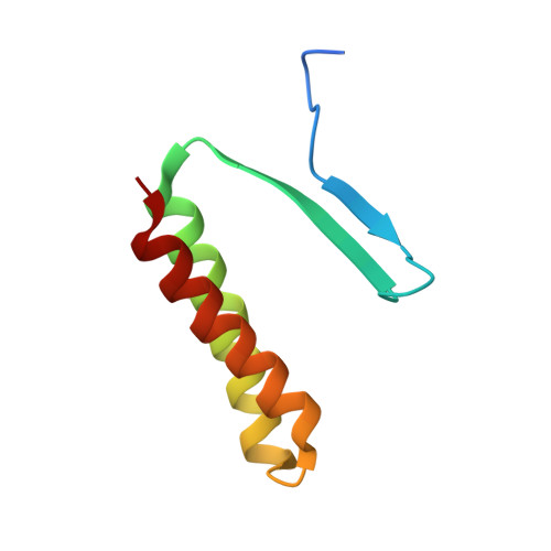
The tetrameric state of p53, p63, and p73 has been considered one of the hallmarks of this protein family. While the DNA binding domain (DBD) is highly conserved among vertebrates and invertebrates, sequences C-terminal to the DBD are highly divergent. In particular, the oligomerization domain (OD) of the p53 forms of the model organisms Caenorhabditis elegans and Drosophila cannot be identified by sequence analysis. Here, we present the solution structures of their ODs and show that they both differ significantly from each other as well as from human p53. CEP-1 contains a composite domain of an OD and a sterile alpha motif (SAM) domain, and forms dimers instead of tetramers. The Dmp53 structure is characterized by an additional N-terminal beta-strand and a C-terminal helix. Truncation analysis in both domains reveals that the additional structural elements are necessary to stabilize the structure of the OD, suggesting a new function for the SAM domain. Furthermore, these structures show a potential path of evolution from an ancestral dimeric form over a tetrameric form, with additional stabilization elements, to the tetramerization domain of mammalian p53.
- Institute of Biophysical Chemistry, Centre for Biomolecular Magnetic Resonance (BMRZ), JW Goethe University of Frankfurt, Frankfurt/Main, Germany.
Organizational Affiliation: