Molecular basis for interprotein complex-dependent effects on the redox properties of amicyanin.
Zhu, Z., Cunane, L.M., Chen, Z., Durley, R.C., Mathews, F.S., Davidson, V.L.(1998) Biochemistry 37: 17128-17136
- PubMed: 9860825
- DOI: https://doi.org/10.1021/bi9817919
- Primary Citation of Related Structures:
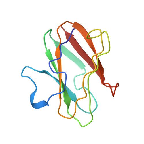
1BXA, 2RAC - PubMed Abstract:
The quinoprotein methylamine dehydrogenase (MADH), type I copper protein amicyanin, and cytochrome c-551i form a complex within which interprotein electron transfer occurs. It was known that complex formation significantly lowered the oxidation-reduction midpoint potential (Em) value of amicyanin, which facilitated an otherwise thermodynamically unfavorable electron transfer to cytochrome c-551i. Structural, mutagenesis, and potentiometric studies have elucidated the basis for this complex-dependent change in redox properties. Positively charged amino acid residues on the surface of amicyanin are known to stabilize complex formation with MADH and influence the ionic strength dependence of complex formation via electrostatic interactions. Altering the charges of these residues by site-directed mutagenesis had no effect on the Em value of amicyanin, ruling out charge neutralization as the basis for the complex-dependent changes in redox properties. The Em value of free amicyanin varies with pH and exhibits a pKa value for the reduced form of 7.5. The crystal structure of reduced amicyanin at pH 4.4 reveals that His95, which serves as a ligand for Cu2+, has rotated by 180 degrees about the Cbeta-Cgamma bond relative to its position in oxidized amicyanin and is no longer in the copper coordination sphere. At pH 7.7, the crystal structure of reduced amicyanin contains an approximately equal distribution of two active-site conformers. One is very similar to the structure of reduced amicyanin at pH 4.4, and the other is very similar to the structure of oxidized amicyanin at pH 4.8. Potentiometric analysis of amicyanin in complex with MADH indicates that its Em value is not pH-dependent from pH 6.5 to 8.5, and exhibits an Em value similar to that of free amicyanin at high pH. The structure of reduced amicyanin at pH 4.4, with His95 protonated and "flipped", was modeled into the structure of the complex of oxidized amicyanin with MADH. This showed that in the complex, the redox-linked pH-dependent rotation of His95 is hindered because it would cause an overlap of van der Waals' radii with residues of MADH. These results demonstrate that protein-protein interactions profoundly affect the redox properties of this type I copper protein by restricting a pH-dependent, redox-linked conformational change of one of the copper ligands.
Organizational Affiliation:
Department of Biochemistry, The University of Mississippi Medical Center, Jackson 39216-4505, USA.