2Q1E

Altered dimer interface decreases stability in an amyloidogenic kappa1 Bence Jones protein.


Experimental Data Snapshot

  • Method: X-RAY DIFFRACTION
  • Resolution: 2.55 Å
  • R-Value Free: 0.206 
  • R-Value Work: 0.165 
  • R-Value Observed: 0.166 

wwPDB Validation   3D Report Full Report


This is version 1.3 of the entry. See complete history


Literature

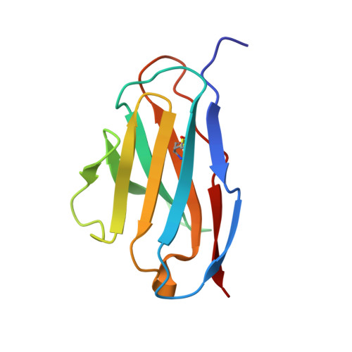
Altered dimer interface decreases stability in an amyloidogenic protein.

Baden, E.M.Owen, B.A.Peterson, F.C.Volkman, B.F.Ramirez-Alvarado, M.Thompson, J.R.

(2008) J Biol Chem 283: 15853-15860

  • DOI: https://doi.org/10.1074/jbc.M705347200
  • Primary Citation of Related Structures:  
    2Q1E, 2Q20

  • PubMed Abstract: 

    Amyloidoses are devastating and currently incurable diseases in which the process of amyloid formation causes fatal cellular and organ damage. The molecular mechanisms underlying amyloidoses are not well known. In this study, we address the structural basis of immunoglobulin light chain amyloidosis, which results from deposition of light chains produced by clonal plasma cells. We compare light chain amyloidosis protein AL-09 to its wild-type counterpart, the kappaI O18/O8 light chain germline. Crystallographic studies indicate that both proteins form dimers. However, AL-09 has an altered dimer interface that is rotated 90 degrees from the kappaI O18/O8 dimer interface. The three non-conservative mutations in AL-09 are located within the dimer interface, consistent with their role in the decreased stability of this amyloidogenic protein. Moreover, AL-09 forms amyloid fibrils more quickly than kappaI O18/O8 in vitro. These results support the notion that the increased stability of the monomer and delayed fibril formation, together with a properly formed dimer, may be protective against amyloidogenesis. This could open a new direction into rational drug design for amyloidogenic proteins.


  • Organizational Affiliation

    Department of Biochemistry and Molecular Biology, College of Medicine, Mayo Clinic, Rochester, Minnesota 55905, USA.


Macromolecules
Find similar proteins by:  (by identity cutoff)  |  3D Structure
Entity ID: 1
MoleculeChains Sequence LengthOrganismDetailsImage
Amyloidogenic immunoglobulin light chain protein AL-09
A, B, C, D
109Homo sapiensMutation(s): 0 
Gene Names: mutant of Vk1 O18/O8 germline
UniProt
Find proteins for A2NI60 (Homo sapiens)
Explore A2NI60 
Go to UniProtKB:  A2NI60
Entity Groups  
Sequence Clusters30% Identity50% Identity70% Identity90% Identity95% Identity100% Identity
UniProt GroupA2NI60
Sequence Annotations
Expand
  • Reference Sequence
Small Molecules
Ligands 1 Unique
IDChains Name / Formula / InChI Key2D Diagram3D Interactions
SO4
Query on SO4

Download Ideal Coordinates CCD File 
E [auth A]SULFATE ION
O4 S
QAOWNCQODCNURD-UHFFFAOYSA-L
Experimental Data & Validation

Experimental Data

  • Method: X-RAY DIFFRACTION
  • Resolution: 2.55 Å
  • R-Value Free: 0.206 
  • R-Value Work: 0.165 
  • R-Value Observed: 0.166 
  • Space Group: P 41 3 2
Unit Cell:
Length ( Å )Angle ( ˚ )
a = 176.054α = 90
b = 176.054β = 90
c = 176.054γ = 90
Software Package:
Software NamePurpose
REFMACrefinement
SBC-Collectdata collection
HKL-3000data reduction
HKL-3000data scaling
PHASERphasing

Structure Validation

View Full Validation Report


Entry History 

Deposition Data

Revision History  (Full details and data files)

  • Version 1.0: 2008-04-08
    Type: Initial release
  • Version 1.1: 2011-07-13
    Changes: Version format compliance
  • Version 1.2: 2017-10-18
    Changes: Refinement description
  • Version 1.3: 2024-04-03
    Changes: Data collection, Database references, Derived calculations, Refinement description