2PRI

BINDING OF 2-DEOXY-GLUCOSE-6-PHOSPHATE TO GLYCOGEN PHOSPHORYLASE B


Experimental Data Snapshot

  • Method: X-RAY DIFFRACTION
  • Resolution: 2.30 Å
  • R-Value Free: 0.246 
  • R-Value Work: 0.192 
  • R-Value Observed: 0.192 

wwPDB Validation   3D Report Full Report


This is version 1.6 of the entry. See complete history


Literature

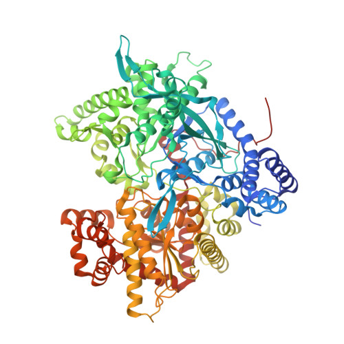
The binding of 2-deoxy-D-glucose 6-phosphate to glycogen phosphorylase b: kinetic and crystallographic studies.

Oikonomakos, N.G.Zographos, S.E.Johnson, L.N.Papageorgiou, A.C.Acharya, K.R.

(1995) J Mol Biol 254: 900-917

  • DOI: https://doi.org/10.1006/jmbi.1995.0665
  • Primary Citation of Related Structures:  
    2PRI

  • PubMed Abstract: 

    Kinetic and crystallographic studies have characterized the effect of 2-deoxy-glucose 6-phosphate on the catalytic and structural properties of glycogen phosphorylase b. Previous work on the binding of glucose 6-phosphate, a potent physiological inhibitor of the enzyme, to T state phosphorylase b in the crystal showed that the inhibitor binds at the allosteric site and induces substantial conformational changes that affect the subunit-subunit interface. The hydrogen-bond from the O-2 hydroxyl of glucose 6-phosphate to the main-chain oxygen of Val40' represents the only hydrogen bond from the sugar to the other subunit, and this interaction appears important for promoting a more "tensed" structure than native T state phosphorylase b. 2-Deoxy-glucose 6-phosphate acts competitively with both the activator AMP and the substrate glucose 1-phosphate, with Ki values of 0.53 mM and 1.23 mM, respectively. The binding of 2-deoxy-glucose 6-phosphate to T state glycogen phosphorylase b in the crystal, has been investigated and the complex phosphorylase b: 2-deoxy-glucose 6-phosphate has been refined to give a crystallographic R factor of 17.3%, for data between 8 A and 2.3 A. 2-Deoxy-glucose 6-phosphate binds at the allosteric site as the a anomer and adopts a different conformation compared to glucose 6-phosphate. The two conformations differ by 160 degrees in the torsion angle about the C-5-C-6 bond. The contacts from the phosphate group are essentially identical to those made by the phosphate of glucose 6-phosphate but the 2-deoxy glucosyl moiety binds in a quite different orientation compared to the glucosyl of glucose 6-phosphate. 2-Deoxy-glucose 6-phosphate can be accommodated in the allosteric site with very little change in the protein, while structural comparisons show that the phosphorylase b: 2-deoxy-glucose 6-phosphate complex structure is overall more similar to a glucose-like complex than to the Glc-6-P complex structure.


  • Organizational Affiliation

    Institute of Biological Research & Biotechnology, National Hellenic Research Foundation, Athens, Greece.


Macromolecules
Find similar proteins by:  (by identity cutoff)  |  3D Structure
Entity ID: 1
MoleculeChains Sequence LengthOrganismDetailsImage
GLYCOGEN PHOSPHORYLASE B842Oryctolagus cuniculusMutation(s): 0 
EC: 2.4.1.1
UniProt
Find proteins for P00489 (Oryctolagus cuniculus)
Explore P00489 
Go to UniProtKB:  P00489
Entity Groups  
Sequence Clusters30% Identity50% Identity70% Identity90% Identity95% Identity100% Identity
UniProt GroupP00489
Sequence Annotations
Expand
  • Reference Sequence
Small Molecules
Ligands 2 Unique
IDChains Name / Formula / InChI Key2D Diagram3D Interactions
PLP
Query on PLP

Download Ideal Coordinates CCD File 
C [auth A]PYRIDOXAL-5'-PHOSPHATE
C8 H10 N O6 P
NGVDGCNFYWLIFO-UHFFFAOYSA-N
D6G
Query on D6G

Download Ideal Coordinates CCD File 
B [auth A]2-deoxy-6-O-phosphono-alpha-D-glucopyranose
C6 H13 O8 P
UQJFZAAGZAYVKZ-ZXXMMSQZSA-N
Binding Affinity Annotations 
IDSourceBinding Affinity
D6G PDBBind:  2PRI Ki: 1.23e+6 (nM) from 1 assay(s)
Binding MOAD:  2PRI Ki: 1.23e+6 (nM) from 1 assay(s)
Experimental Data & Validation

Experimental Data

  • Method: X-RAY DIFFRACTION
  • Resolution: 2.30 Å
  • R-Value Free: 0.246 
  • R-Value Work: 0.192 
  • R-Value Observed: 0.192 
  • Space Group: P 43 21 2
Unit Cell:
Length ( Å )Angle ( ˚ )
a = 128.5α = 90
b = 128.5β = 90
c = 116.3γ = 90
Software Package:
Software NamePurpose
X-PLORrefinement
XDSdata reduction

Structure Validation

View Full Validation Report


Entry History 

Deposition Data

Revision History  (Full details and data files)

  • Version 1.0: 1998-12-14
    Type: Initial release
  • Version 1.1: 2008-04-26
    Changes: Version format compliance
  • Version 1.2: 2011-07-13
    Changes: Derived calculations, Version format compliance
  • Version 1.3: 2012-08-29
    Changes: Non-polymer description
  • Version 1.4: 2018-03-07
    Changes: Data collection
  • Version 1.5: 2020-07-29
    Type: Remediation
    Reason: Carbohydrate remediation
    Changes: Data collection, Derived calculations, Structure summary
  • Version 1.6: 2023-08-30
    Changes: Data collection, Database references, Refinement description, Structure summary