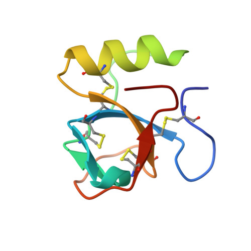
Crystal Structures of Hydrophobin HFBII in the Presence of Detergent Implicate the Formation of Fibrils and Monolayer Films.
Kallio, J.M., Linder, M.B., Rouvinen, J.(2007) J Biological Chem 282: 28733-28739
- PubMed: 17636262
- DOI: https://doi.org/10.1074/jbc.M704238200
- Primary Citation of Related Structures:
2PL6, 2PL7 - PubMed Abstract:
Hydrophobins are small, amphiphilic proteins secreted by filamentous fungi. Their functionality arises from a patch of hydrophobic residues on the protein surface. Spontaneous self-assembly of hydrophobins leads to the formation of an amphiphilic layer that remarkably reduces the surface tension of water. We have determined by x-ray diffraction two new crystal structures of Trichoderma reesei hydrophobin HFBII in the presence of a detergent. The monoclinic crystal structure (2.2A resolution, R = 22, R(free) = 28) is composed of layers of hydrophobin molecules where the hydrophobic surface areas of the molecules are aligned within the layer. Viewed perpendicular to the aligned hydrophobic surface areas, the molecules in the layer pack together to form six-membered rings, thus leaving small pores in the layer. Similar packing has been observed in the atomic force microscopy images of the self-assembled layers of class II hydrophobin, indicating that the crystal structure resembles that of natural hydrophobin film. The orthorhombic crystal structure (1.0 A resolution, R = 13, R(free) = 15) is composed of fiber-like arrays of protein molecules. Rodlet structures have been observed on amphiphilic layers formed by class I hydrophobins; fibrils of class II hydrophobins appear by vigorous shaking. We propose that the structure of the fibrils and/or rodlets is similar to that observed in the crystal structure.
- Department of Chemistry, University of Joensuu, P. O. Box 111, 80101 Joensuu, Finland. Electronic address: johanna.kallio@joensuu.fi.
Organizational Affiliation: