2OHF

Crystal structure of human OLA1 in complex with AMPPCP


Experimental Data Snapshot

  • Method: X-RAY DIFFRACTION
  • Resolution: 2.70 Å
  • R-Value Free: 0.288 
  • R-Value Work: 0.230 
  • R-Value Observed: 0.233 

wwPDB Validation   3D Report Full Report


Ligand Structure Quality Assessment 


This is version 1.4 of the entry. See complete history


Literature

Human OLA1 defines an ATPase subfamily in the Obg family of GTP-binding proteins

Koller-Eichhorn, R.Marquardt, T.Gail, R.Wittinghofer, A.Kostrewa, D.Kutay, U.Kambach, C.

(2007) J Biol Chem 282: 19928-19937

  • DOI: https://doi.org/10.1074/jbc.M700541200
  • Primary Citation of Related Structures:  
    2OHF

  • PubMed Abstract: 

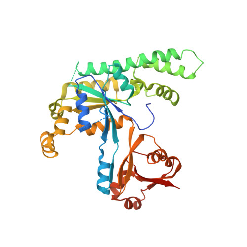
    Purine nucleotide-binding proteins build the large family of P-loop GTPases and related ATPases, which perform essential functions in all kingdoms of life. The Obg family comprises a group of ancient GTPases belonging to the TRAFAC (for translation factors) class and can be subdivided into several distinct protein subfamilies. The founding member of one of these subfamilies is the bacterial P-loop NTPase YchF, which had so far been assumed to act as GTPase. We have biochemically characterized the human homologue of YchF and found that it binds and hydrolyzes ATP more efficiently than GTP. For this reason, we have termed the protein hOLA1, for human Obg-like ATPase 1. Further biochemical characterization of YchF proteins from different species revealed that ATPase activity is a general but previously missed feature of the YchF subfamily of Obg-like GTPases. To explain ATP specificity of hOLA1, we have solved the x-ray structure of hOLA1 bound to the nonhydrolyzable ATP analogue AMPPCP. Our structural data help to explain the altered nucleotide specificity of YchF homologues and identify the Ola1/YchF subfamily of the Obg-related NTPases as an exceptional example of a single protein subfamily, which has evolved altered nucleotide specificity within a distinct protein family of GTPases.


  • Organizational Affiliation

    Institute of Biochemistry, ETH Zurich, 8093 Zurich, Switzerland.


Macromolecules
Find similar proteins by:  (by identity cutoff)  |  3D Structure
Entity ID: 1
MoleculeChains Sequence LengthOrganismDetailsImage
GTP-binding protein 9396Homo sapiensMutation(s): 0 
Gene Names: GTPBP9
UniProt & NIH Common Fund Data Resources
Find proteins for Q9NTK5 (Homo sapiens)
Explore Q9NTK5 
Go to UniProtKB:  Q9NTK5
PHAROS:  Q9NTK5
GTEx:  ENSG00000138430 
Entity Groups  
Sequence Clusters30% Identity50% Identity70% Identity90% Identity95% Identity100% Identity
UniProt GroupQ9NTK5
Sequence Annotations
Expand
  • Reference Sequence
Small Molecules
Ligands 1 Unique
IDChains Name / Formula / InChI Key2D Diagram3D Interactions
ACP
Query on ACP

Download Ideal Coordinates CCD File 
B [auth A]PHOSPHOMETHYLPHOSPHONIC ACID ADENYLATE ESTER
C11 H18 N5 O12 P3
UFZTZBNSLXELAL-IOSLPCCCSA-N
Experimental Data & Validation

Experimental Data

  • Method: X-RAY DIFFRACTION
  • Resolution: 2.70 Å
  • R-Value Free: 0.288 
  • R-Value Work: 0.230 
  • R-Value Observed: 0.233 
  • Space Group: C 2 2 21
Unit Cell:
Length ( Å )Angle ( ˚ )
a = 141.253α = 90
b = 159.831β = 90
c = 55.129γ = 90
Software Package:
Software NamePurpose
MAR345data collection
MOLREPphasing
REFMACrefinement
XDSdata reduction
XSCALEdata scaling

Structure Validation

View Full Validation Report


Ligand Structure Quality Assessment 


Entry History 

Deposition Data

Revision History  (Full details and data files)

  • Version 1.0: 2007-04-24
    Type: Initial release
  • Version 1.1: 2008-05-01
    Changes: Version format compliance
  • Version 1.2: 2011-07-13
    Changes: Advisory, Version format compliance
  • Version 1.3: 2017-10-18
    Changes: Refinement description
  • Version 1.4: 2023-08-30
    Changes: Data collection, Database references, Derived calculations, Refinement description