2MV1

Solution NMR structure of Human Relaxin-2


Experimental Data Snapshot

  • Method: SOLUTION NMR
  • Conformers Calculated: 50 
  • Conformers Submitted: 20 
  • Selection Criteria: structures with the lowest energy 

wwPDB Validation   3D Report Full Report


This is version 2.0 of the entry. See complete history


Literature

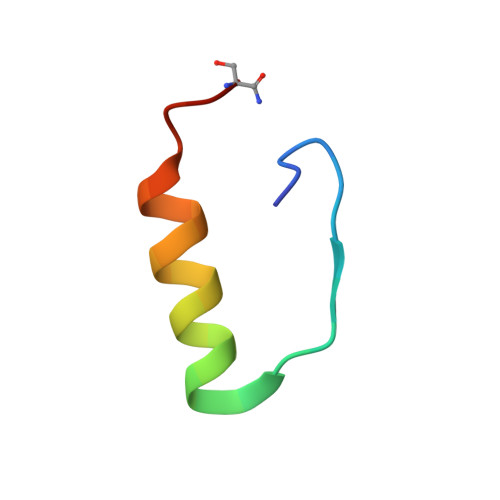
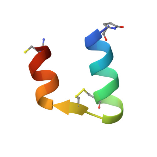
Solution structure, aggregation behavior, and flexibility of human relaxin-2.

Haugaard-Kedstrom, L.M.Hossain, M.A.Daly, N.L.Bathgate, R.A.Rinderknecht, E.Wade, J.D.Craik, D.J.Rosengren, K.J.

(2015) ACS Chem Biol 10: 891-900

  • DOI: https://doi.org/10.1021/cb500918v
  • Primary Citation of Related Structures:  
    2MV1

  • PubMed Abstract: 

    Relaxin is a member of the relaxin/insulin peptide hormone superfamily and is characterized by a two-chain structure constrained by three disulfide bonds. Relaxin is a pleiotropic hormone and involved in a number of physiological and pathogenic processes, including collagen and cardiovascular regulation and tissue remodelling during pregnancy and cancer. Crystallographic and ultracentrifugation experiments have revealed that the human form of relaxin, H2 relaxin, self-associates into dimers, but the significance of this is poorly understood. Here, we present the NMR structure of a monomeric, amidated form of H2 relaxin and compare its features and behavior in solution to those of native H2 relaxin. The overall structure of H2 relaxin is retained in the monomeric form. H2 relaxin amide is fully active at the relaxin receptor RXFP1 and thus dimerization is not required for biological activity. Analysis of NMR chemical shifts and relaxation parameters identified internal motion in H2 relaxin at the pico-nanosecond and milli-microsecond time scales, which is commonly seen in other relaxin and insulin peptides and might be related to function.


  • Organizational Affiliation

    □School of Natural Sciences, Linnaeus University, SE-391 82 Kalmar, Sweden.


Macromolecules
Find similar proteins by:  (by identity cutoff)  |  3D Structure
Entity ID: 1
MoleculeChains Sequence LengthOrganismDetailsImage
Relaxin B chainA [auth B]30Homo sapiensMutation(s): 1 
UniProt & NIH Common Fund Data Resources
Find proteins for P04090 (Homo sapiens)
Explore P04090 
Go to UniProtKB:  P04090
PHAROS:  P04090
GTEx:  ENSG00000107014 
Entity Groups  
Sequence Clusters30% Identity50% Identity70% Identity90% Identity95% Identity100% Identity
UniProt GroupP04090
Sequence Annotations
Expand
  • Reference Sequence

Find similar proteins by:  Sequence   |   3D Structure  

Entity ID: 2
MoleculeChains Sequence LengthOrganismDetailsImage
Relaxin A chainB [auth A]25Homo sapiensMutation(s): 1 
UniProt & NIH Common Fund Data Resources
Find proteins for P04090 (Homo sapiens)
Explore P04090 
Go to UniProtKB:  P04090
PHAROS:  P04090
GTEx:  ENSG00000107014 
Entity Groups  
Sequence Clusters30% Identity50% Identity70% Identity90% Identity95% Identity100% Identity
UniProt GroupP04090
Sequence Annotations
Expand
  • Reference Sequence
Small Molecules
Modified Residues  1 Unique
IDChains TypeFormula2D DiagramParent
PCA
Query on PCA
B [auth A]L-PEPTIDE LINKINGC5 H7 N O3GLN
Experimental Data & Validation

Experimental Data

  • Method: SOLUTION NMR
  • Conformers Calculated: 50 
  • Conformers Submitted: 20 
  • Selection Criteria: structures with the lowest energy 

Structure Validation

View Full Validation Report


Entry History 

Deposition Data

Revision History  (Full details and data files)

  • Version 1.0: 2015-02-04
    Type: Initial release
  • Version 1.1: 2015-04-22
    Changes: Database references
  • Version 2.0: 2019-12-25
    Changes: Data collection, Database references, Derived calculations, Polymer sequence