Hydrophobic matching controls the tilt and stability of the dimeric platelet-derived growth factor receptor (PDGFR) beta transmembrane segment.
Muhle-Goll, C., Hoffmann, S., Afonin, S., Grage, S.L., Polyansky, A.A., Windisch, D., Zeitler, M., Burck, J., Ulrich, A.S.(2012) J Biol Chem 287: 26178-26186
- PubMed: 22619173
- DOI: https://doi.org/10.1074/jbc.M111.325555
- Primary Citation of Related Structures:
2L6W - PubMed Abstract:
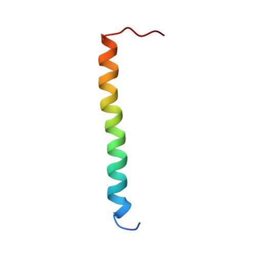
The platelet-derived growth factor receptor β is a member of the cell surface receptor tyrosine kinase family and dimerizes upon activation. We determined the structure of the transmembrane segment in dodecylphosphocholine micelles by liquid-state NMR and found that it forms a stable left-handed helical dimer. Solid-state NMR and oriented circular dichroism were used to measure the tilt angle of the helical segments in macroscopically aligned model membranes with different acyl chain lengths. Both methods showed that decreasing bilayer thickness (DEPC-POPC-DMPC) led to an increase in the helix tilt angle from 10° to 30° with respect to the bilayer normal. At the same time, reconstitution of the comparatively long hydrophobic segment became less effective, eventually resulting in complete protein aggregation in the short-chain lipid DLPC. Unrestrained molecular dynamics simulations of the dimer were carried out in explicit lipid bilayers (DEPC, POPC, DMPC, sphingomyelin), confirming the observed dependence of the helix tilt angle on bilayer thickness. Notably, molecular dynamics revealed that the left-handed dimer gets tilted en bloc, whereas conformational transitions to alternative (e.g. right-handed dimeric) states were not supported. The experimental data along with the simulation results demonstrate a pronounced interplay between the platelet-directed growth factor receptor β transmembrane segment and the bilayer thickness. The effect of hydrophobic mismatch might play a key role in the redistribution and activation of the receptor within different lipid microdomains of the plasma membrane in vivo.
Organizational Affiliation:
Institute for Biological Interfaces (IBG-2), Karlsruhe Institute of Technology, P. O. Box 3640, 76021 Karlsruhe, Germany.