2J4O

Structure of TAB1


Experimental Data Snapshot

  • Method: X-RAY DIFFRACTION
  • Resolution: 2.25 Å
  • R-Value Free: 0.236 
  • R-Value Work: 0.228 
  • R-Value Observed: 0.228 

wwPDB Validation   3D Report Full Report


This is version 1.3 of the entry. See complete history


Literature

Tak1-Binding Protein 1 is a Pseudophosphatase.

Conner, S.H.Kular, G.Peggie, M.Shepherd, S.Schuttelkopf, A.W.Cohen, P.Van Aalten, D.M.F.

(2006) Biochem J 399: 427

  • DOI: https://doi.org/10.1042/BJ20061077
  • Primary Citation of Related Structures:  
    2J4O

  • PubMed Abstract: 

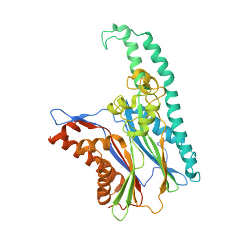
    TAB1 [TAK1 (transforming growth factor-beta-activated kinase 1)-binding protein 1] is one of the regulatory subunits of TAK1, a protein kinase that lies at the head of three pro-inflammatory kinase cascades. In the current study we report the crystal structure of the N-terminal domain of TAB1. Surprisingly, TAB1 possesses a fold closely related to that of the PPM (Mg2+- or Mn2+-dependent protein phosphatase) family as demonstrated by the close structural similarity with protein phosphatase 2C alpha. However, we were unable to detect any phosphatase activity for TAB1 using a phosphopeptide or p-nitrophenyl phosphate as substrate. Although the overall protein phosphatase 2C alpha fold is conserved in TAB1, detailed structural analyses and mutagenesis studies show that several key residues required for dual metal-binding and catalysis are not present in TAB1, although binding of a single metal is supported by soaking experiments with manganese and isothermal titration calorimetry. Thus, it appears that TAB1 is a 'pseudophosphatase', possibly binding to and regulating accessibility of phosphorylated residues on substrates downstream of TAK1 or on the TAK1 complex itself.


  • Organizational Affiliation

    MRC Protein Phosphorylation Unit, School of Life Sciences, University of Dundee, Dundee DD1 5EH, Scotland, UK.


Macromolecules
Find similar proteins by:  (by identity cutoff)  |  3D Structure
Entity ID: 1
MoleculeChains Sequence LengthOrganismDetailsImage
MITOGEN-ACTIVATED PROTEIN KINASE KINASE KINASE 7-INTERACTING PROTEIN 1401Homo sapiensMutation(s): 0 
UniProt & NIH Common Fund Data Resources
Find proteins for Q15750 (Homo sapiens)
Explore Q15750 
Go to UniProtKB:  Q15750
PHAROS:  Q15750
GTEx:  ENSG00000100324 
Entity Groups  
Sequence Clusters30% Identity50% Identity70% Identity90% Identity95% Identity100% Identity
UniProt GroupQ15750
Sequence Annotations
Expand
  • Reference Sequence
Experimental Data & Validation

Experimental Data

  • Method: X-RAY DIFFRACTION
  • Resolution: 2.25 Å
  • R-Value Free: 0.236 
  • R-Value Work: 0.228 
  • R-Value Observed: 0.228 
  • Space Group: P 3 2 1
Unit Cell:
Length ( Å )Angle ( ˚ )
a = 141.961α = 90
b = 141.961β = 90
c = 65.905γ = 120
Software Package:
Software NamePurpose
CNSrefinement
DENZOdata reduction
SCALEPACKdata scaling
SHELXphasing

Structure Validation

View Full Validation Report


Entry History 

Deposition Data

Revision History  (Full details and data files)

  • Version 1.0: 2006-09-04
    Type: Initial release
  • Version 1.1: 2011-05-08
    Changes: Version format compliance
  • Version 1.2: 2011-07-13
    Changes: Version format compliance
  • Version 1.3: 2019-03-06
    Changes: Data collection, Experimental preparation, Other