2IXM

Structure of human PTPA


Experimental Data Snapshot

  • Method: X-RAY DIFFRACTION
  • Resolution: 1.50 Å
  • R-Value Free: 0.221 
  • R-Value Work: 0.202 
  • R-Value Observed: 0.203 

wwPDB Validation   3D Report Full Report


This is version 1.4 of the entry. See complete history


Literature

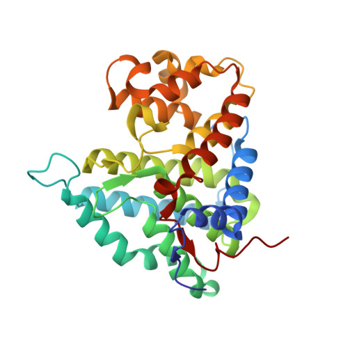
Crystal Structure of the Pp2A Phosphatase Activator: Implications for its Pp2A-Specific Ppiase Activity

Leulliot, N.Vicentini, G.Jordens, J.Quevillon-Cheruel, S.Schiltz, M.Barford, D.Van Tilbeurgh, H.Goris, J.

(2006) Mol Cell 23: 413

  • DOI: https://doi.org/10.1016/j.molcel.2006.07.008
  • Primary Citation of Related Structures:  
    2IXM, 2IXN, 2IXO, 2IXP

  • PubMed Abstract: 

    PTPA, an essential and specific activator of protein phosphatase 2A (PP2A), functions as a peptidyl prolyl isomerase (PPIase). We present here the crystal structures of human PTPA and of the two yeast orthologs (Ypa1 and Ypa2), revealing an all alpha-helical protein fold that is radically different from other PPIases. The protein is organized into two domains separated by a groove lined by highly conserved residues. To understand the molecular mechanism of PTPA activity, Ypa1 was cocrystallized with a proline-containing PPIase peptide substrate. In the complex, the peptide binds at the interface of a peptide-induced dimer interface. Conserved residues of the interdomain groove contribute to the peptide binding site and dimer interface. Structure-guided mutational studies showed that in vivo PTPA activity is influenced by mutations on the surface of the peptide binding pocket, the same mutations that also influenced the in vitro activation of PP2Ai and PPIase activity.


  • Organizational Affiliation

    Institut de Biochimie et de Biophysique Moléculaire et Cellulaire, UMR8619, Bât 430, Université de Paris-Sud, 91405 Orsay Cedex, France.


Macromolecules
Find similar proteins by:  (by identity cutoff)  |  3D Structure
Entity ID: 1
MoleculeChains Sequence LengthOrganismDetailsImage
SERINE/THREONINE-PROTEIN PHOSPHATASE 2A REGULATORY SUBUNIT B'303Homo sapiensMutation(s): 0 
UniProt & NIH Common Fund Data Resources
Find proteins for Q15257 (Homo sapiens)
Explore Q15257 
Go to UniProtKB:  Q15257
PHAROS:  Q15257
GTEx:  ENSG00000119383 
Entity Groups  
Sequence Clusters30% Identity50% Identity70% Identity90% Identity95% Identity100% Identity
UniProt GroupQ15257
Sequence Annotations
Expand
  • Reference Sequence
Experimental Data & Validation

Experimental Data

  • Method: X-RAY DIFFRACTION
  • Resolution: 1.50 Å
  • R-Value Free: 0.221 
  • R-Value Work: 0.202 
  • R-Value Observed: 0.203 
  • Space Group: P 21 21 21
Unit Cell:
Length ( Å )Angle ( ˚ )
a = 48.243α = 90
b = 70.41β = 90
c = 95.534γ = 90
Software Package:
Software NamePurpose
REFMACrefinement
MOSFLMdata reduction
SCALAdata scaling
SHELXphasing

Structure Validation

View Full Validation Report


Entry History 

Deposition Data

Revision History  (Full details and data files)

  • Version 1.0: 2006-07-11
    Type: Initial release
  • Version 1.1: 2011-05-08
    Changes: Version format compliance
  • Version 1.2: 2011-07-13
    Changes: Version format compliance
  • Version 1.3: 2019-05-08
    Changes: Data collection, Experimental preparation, Other
  • Version 1.4: 2019-05-15
    Changes: Data collection, Experimental preparation