2IER

Crystal Structure of Aquifex aeolicus LpxC Complexed with Uridine 5'-Diphosphate


Experimental Data Snapshot

  • Method: X-RAY DIFFRACTION
  • Resolution: 2.70 Å
  • R-Value Free: 0.241 
  • R-Value Work: 0.213 

wwPDB Validation   3D Report Full Report


Ligand Structure Quality Assessment 


This is version 1.4 of the entry. See complete history


Literature

Binding of Uridine 5-Diphosphate in the Basic Patch of the Zinc Metalloenzyme Deacetylase LpxC and Implications for Substrate Binding

Gennadios, H.A.Christianson, D.W.

(2006) Biochemistry 45: 15216-15223

  • DOI: https://doi.org/10.1021/bi0619021
  • Primary Citation of Related Structures:  
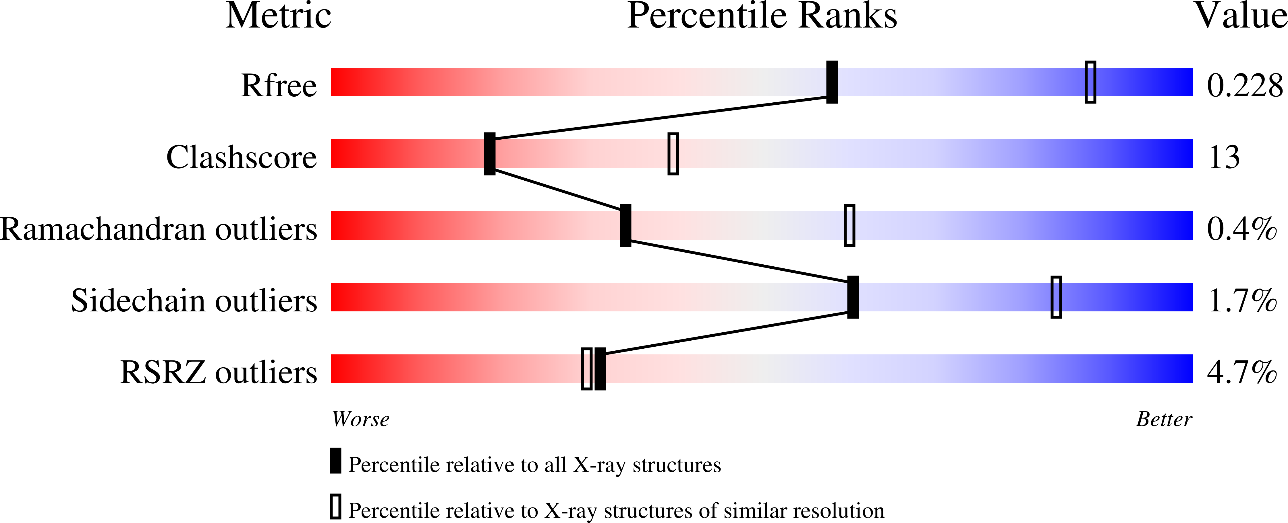
    2IER, 2IES

  • PubMed Abstract: 

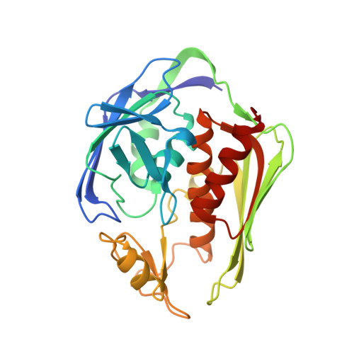
    LpxC is a zinc metalloenzyme that catalyzes the first committed step in the biosynthesis of lipid A, a vital component of the outer membrane of Gram-negative bacteria. Accordingly, the inhibition of LpxC is an attractive strategy for the treatment of Gram-negative bacterial infections. Here, we report the 2.7 A resolution X-ray crystal structure of LpxC from Aquifex aeolicus complexed with uridine 5'-diphosphate (UDP), and the 3.1 A resolution structure of LpxC complexed with pyrophosphate. The X-ray crystal structure of the LpxC-UDP complex provides the first view of interactions likely to be exploited by the substrate UDP group in the "basic patch" of the active site. The diphosphate group of UDP makes hydrogen bond interactions with strictly conserved residue K239 as well as solvent molecules. The ribose moiety of UDP interacts with partially conserved residue E197. The UDP uracil group hydrogen bonds with both the backbone NH group and the backbone carbonyl group of E160, and with the backbone NH group of K162 through an intervening water molecule. Finally, the alpha-phosphate and uracil groups of UDP interact with R143 and R262 through intervening water molecules. The structure of LpxC complexed with pyrophosphate reveals generally similar intermolecular interactions in the basic patch. Unexpectedly, diphosphate binding in both complexes is accompanied by coordination to an additional zinc ion, resulting in the identification of a new metal-binding site termed the E-site. The structures of the LpxC-UDP and LpxC-pyrophosphate complexes provide new insights with regard to substrate recognition in the basic patch and metal ion coordination in the active site of LpxC.


  • Organizational Affiliation

    Roy and Diana Vagelos Laboratories, Department of Chemistry, University of Pennsylvania, Philadelphia, Pennsylvania 19104-6323.


Macromolecules
Find similar proteins by:  (by identity cutoff)  |  3D Structure
Entity ID: 1
MoleculeChains Sequence LengthOrganismDetailsImage
UDP-3-O-[3-hydroxymyristoyl] N-acetylglucosamine deacetylase
A, B
271Aquifex aeolicusMutation(s): 1 
Gene Names: lpxc
EC: 3.5.1
UniProt
Find proteins for O67648 (Aquifex aeolicus (strain VF5))
Explore O67648 
Go to UniProtKB:  O67648
Entity Groups  
Sequence Clusters30% Identity50% Identity70% Identity90% Identity95% Identity100% Identity
UniProt GroupO67648
Sequence Annotations
Expand
  • Reference Sequence
Small Molecules
Ligands 4 Unique
IDChains Name / Formula / InChI Key2D Diagram3D Interactions
UDP
Query on UDP

Download Ideal Coordinates CCD File 
H [auth A],
O [auth B]
URIDINE-5'-DIPHOSPHATE
C9 H14 N2 O12 P2
XCCTYIAWTASOJW-XVFCMESISA-N
PLM
Query on PLM

Download Ideal Coordinates CCD File 
I [auth A],
P [auth B]
PALMITIC ACID
C16 H32 O2
IPCSVZSSVZVIGE-UHFFFAOYSA-N
GOL
Query on GOL

Download Ideal Coordinates CCD File 
J [auth A]GLYCEROL
C3 H8 O3
PEDCQBHIVMGVHV-UHFFFAOYSA-N
ZN
Query on ZN

Download Ideal Coordinates CCD File 
C [auth A]
D [auth A]
E [auth A]
F [auth A]
G [auth A]
C [auth A],
D [auth A],
E [auth A],
F [auth A],
G [auth A],
K [auth B],
L [auth B],
M [auth B],
N [auth B]
ZINC ION
Zn
PTFCDOFLOPIGGS-UHFFFAOYSA-N
Experimental Data & Validation

Experimental Data

  • Method: X-RAY DIFFRACTION
  • Resolution: 2.70 Å
  • R-Value Free: 0.241 
  • R-Value Work: 0.213 
  • Space Group: P 61
Unit Cell:
Length ( Å )Angle ( ˚ )
a = 101.206α = 90
b = 101.206β = 90
c = 122.24γ = 120
Software Package:
Software NamePurpose
CBASSdata collection
AMoREphasing
CNSrefinement
HKL-2000data reduction
HKL-2000data scaling

Structure Validation

View Full Validation Report


Ligand Structure Quality Assessment 


Entry History 

Deposition Data

Revision History  (Full details and data files)

  • Version 1.0: 2007-01-09
    Type: Initial release
  • Version 1.1: 2008-05-01
    Changes: Version format compliance
  • Version 1.2: 2011-07-13
    Changes: Non-polymer description, Version format compliance
  • Version 1.3: 2021-10-20
    Changes: Database references, Derived calculations
  • Version 1.4: 2023-08-30
    Changes: Data collection, Refinement description