2IEI

Crystal structure of rabbit muscle glycogen phosphorylase in complex with 3,4-dihydro-2-quinolone


Experimental Data Snapshot

  • Method: X-RAY DIFFRACTION
  • Resolution: 1.91 Å
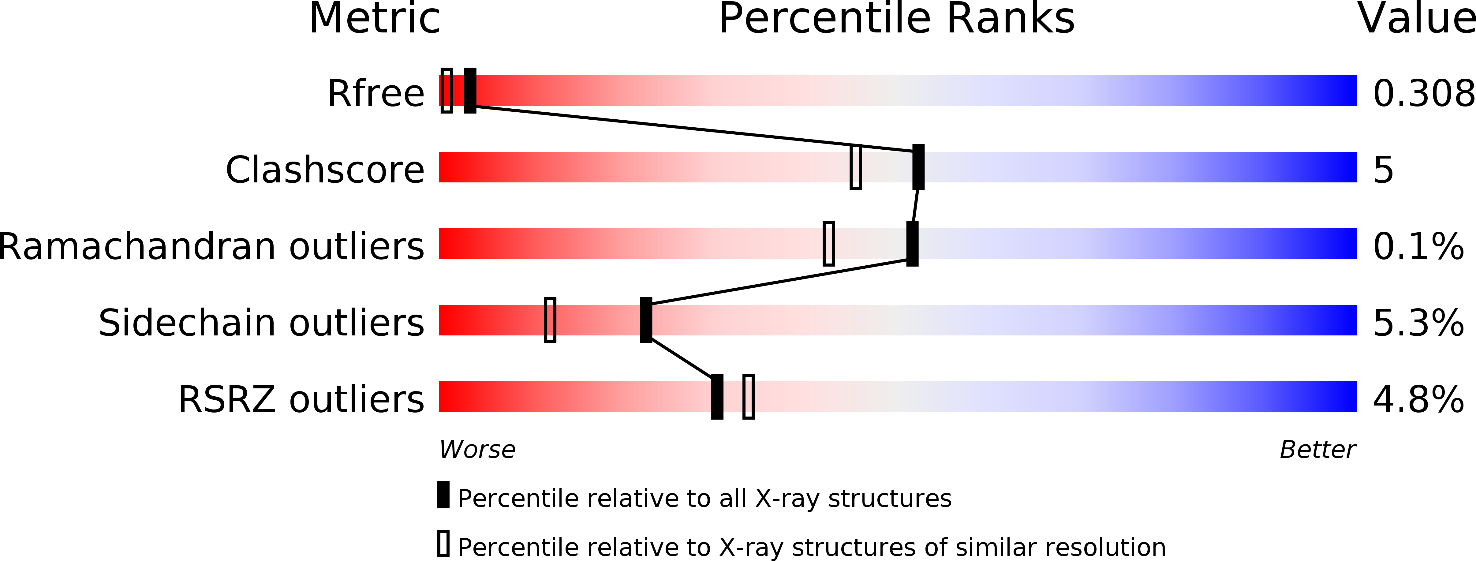
  • R-Value Free: 0.297 
  • R-Value Work: 0.255 
  • R-Value Observed: 0.257 

wwPDB Validation   3D Report Full Report


Ligand Structure Quality Assessment 


This is version 1.2 of the entry. See complete history


Literature

Development of potent, orally active 1-substituted-3,4-dihydro-2-quinolone glycogen phosphorylase inhibitors.

Birch, A.M.Kenny, P.W.Oikonomakos, N.G.Otterbein, L.Schofield, P.Whittamore, P.R.Whalley, D.P.

(2007) Bioorg Med Chem Lett 17: 394-399

  • DOI: https://doi.org/10.1016/j.bmcl.2006.10.037
  • Primary Citation of Related Structures:  
    2IEG, 2IEI

  • PubMed Abstract: 

    A series of substituted 3,4-dihydro-2-quinolone glycogen phosphorylase inhibitors, which have potential as antidiabetic agents, is described. Initial members of the series showed good enzyme inhibitory potency but poor physical properties. Optimisation of the 1-substituent led to 2,3-dihydroxypropyl compounds which showed good in vitro potency and improved physical properties, together with good DMPK profiles and acute in vivo efficacy in a rat model. X-ray crystallographic data are presented, showing an unexpected variety of binding orientations at the dimer interface site.


  • Organizational Affiliation

    AstraZeneca, Mereside, Alderley Park, Macclesfield, Cheshire SK10 4TG, UK. alan.birch@astrazeneca.com


Macromolecules
Find similar proteins by:  (by identity cutoff)  |  3D Structure
Entity ID: 1
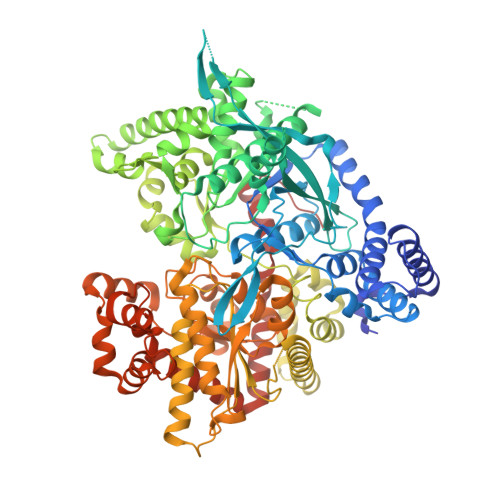
MoleculeChains Sequence LengthOrganismDetailsImage
Glycogen phosphorylase, muscle form
A, B
842Oryctolagus cuniculusMutation(s): 0 
EC: 2.4.1.1
UniProt
Find proteins for P00489 (Oryctolagus cuniculus)
Explore P00489 
Go to UniProtKB:  P00489
Entity Groups  
Sequence Clusters30% Identity50% Identity70% Identity90% Identity95% Identity100% Identity
UniProt GroupP00489
Sequence Annotations
Expand
  • Reference Sequence
Small Molecules
Ligands 2 Unique
IDChains Name / Formula / InChI Key2D Diagram3D Interactions
FRX
Query on FRX

Download Ideal Coordinates CCD File 
D [auth A],
F [auth B]
(S)-2-CHLORO-N-(1-(2-(2-HYDROXYETHYLAMINO)-2-OXOETHYL)-2-OXO-1,2,3,4-TETRAHYDROQUINOLIN-3-YL)-6H-THIENO[2,3-B]PYRROLE-5-CARBOXAMIDE
C20 H19 Cl N4 O4 S
VUKPNWLGSLOHIF-AWEZNQCLSA-N
PLR
Query on PLR

Download Ideal Coordinates CCD File 
C [auth A],
E [auth B]
(5-HYDROXY-4,6-DIMETHYLPYRIDIN-3-YL)METHYL DIHYDROGEN PHOSPHATE
C8 H12 N O5 P
RBCOYOYDYNXAFA-UHFFFAOYSA-N
Binding Affinity Annotations 
IDSourceBinding Affinity
FRX Binding MOAD:  2IEI IC50: 121 (nM) from 1 assay(s)
Experimental Data & Validation

Experimental Data

  • Method: X-RAY DIFFRACTION
  • Resolution: 1.91 Å
  • R-Value Free: 0.297 
  • R-Value Work: 0.255 
  • R-Value Observed: 0.257 
  • Space Group: P 21 21 21
Unit Cell:
Length ( Å )Angle ( ˚ )
a = 114.402α = 90
b = 125.344β = 90
c = 129.309γ = 90
Software Package:
Software NamePurpose
REFMACrefinement
ADSCdata collection
MOSFLMdata reduction
CCP4data scaling
AMoREphasing

Structure Validation

View Full Validation Report


Ligand Structure Quality Assessment 


Entry History 

Revision History  (Full details and data files)

  • Version 1.0: 2006-12-26
    Type: Initial release
  • Version 1.1: 2008-05-01
    Changes: Version format compliance
  • Version 1.2: 2011-07-13
    Changes: Version format compliance