


Crystal structure of the uridine phosphorylase from Salmonella typhimurium in complex with thymine and phosphate ion at 1.70A resolution
Timofeev, V.I., Gabdulkhakov, A.G., Mikhailov, A.M., Dontsova, M.V., Voelter, W., Betzel, C.To be published.
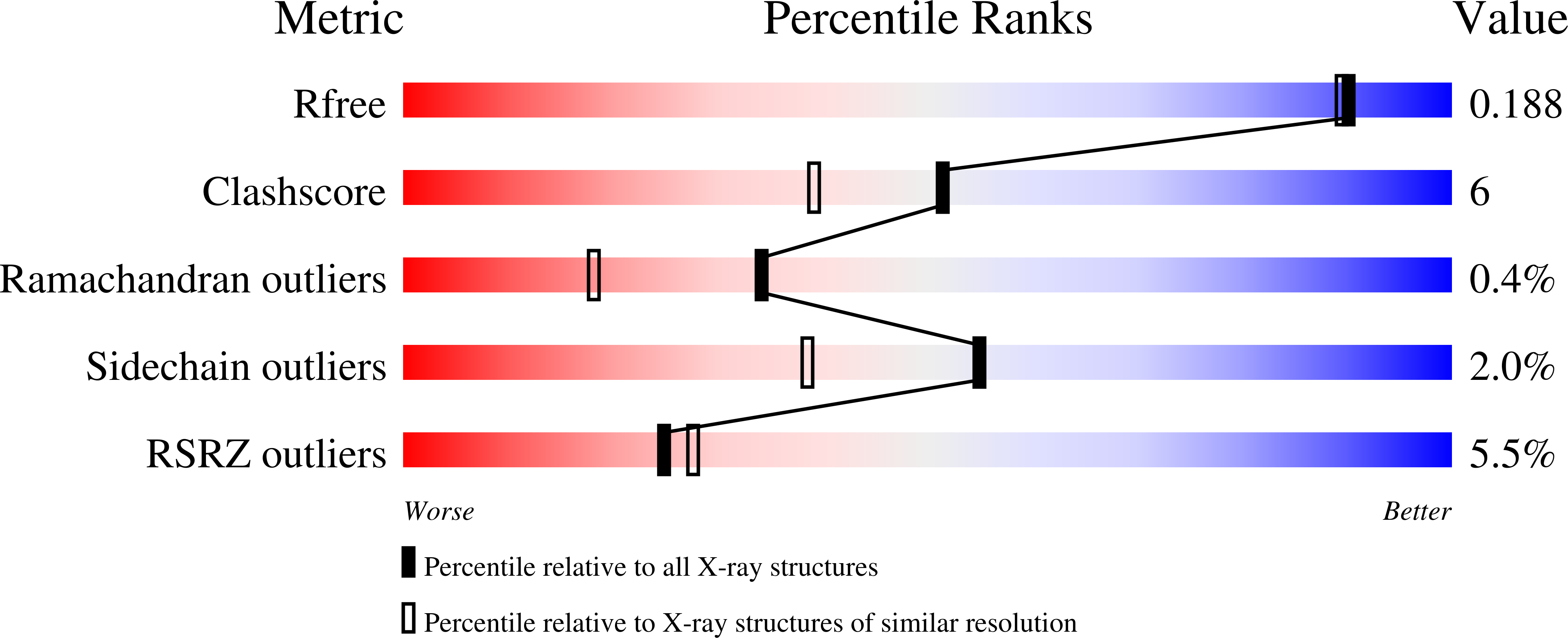
Experimental Data Snapshot
wwPDB Validation 3D Report Full Report
Entity ID: 1 | |||||
|---|---|---|---|---|---|
| Molecule | Chains | Sequence Length | Organism | Details | Image |
| Uridine phosphorylase | 253 | Salmonella enterica subsp. enterica serovar Typhimurium str. LT2 | Mutation(s): 0 Gene Names: UDP EC: 2.4.2.3 |  | |
UniProt | |||||
Find proteins for P0A1F6 (Salmonella typhimurium (strain LT2 / SGSC1412 / ATCC 700720)) Explore P0A1F6 Go to UniProtKB: P0A1F6 | |||||
Entity Groups | |||||
| Sequence Clusters | 30% Identity50% Identity70% Identity90% Identity95% Identity100% Identity | ||||
| UniProt Group | P0A1F6 | ||||
Sequence AnnotationsExpand | |||||
| |||||
| Ligands 4 Unique | |||||
|---|---|---|---|---|---|
| ID | Chains | Name / Formula / InChI Key | 2D Diagram | 3D Interactions | |
| 1PE Query on 1PE | M [auth B] | PENTAETHYLENE GLYCOL C10 H22 O6 JLFNLZLINWHATN-UHFFFAOYSA-N |  | ||
| TDR Query on TDR | I [auth A] L [auth B] P [auth C] S [auth D] U [auth E] | THYMINE C5 H6 N2 O2 RWQNBRDOKXIBIV-UHFFFAOYSA-N |  | ||
| PO4 Query on PO4 | G [auth A] H [auth A] K [auth B] O [auth C] R [auth D] | PHOSPHATE ION O4 P NBIIXXVUZAFLBC-UHFFFAOYSA-K |  | ||
| GOL Query on GOL | J [auth A], N [auth B], Q [auth C], V [auth E], Z [auth F] | GLYCEROL C3 H8 O3 PEDCQBHIVMGVHV-UHFFFAOYSA-N |  | ||
| Length ( Å ) | Angle ( ˚ ) |
|---|---|
| a = 88.2 | α = 90 |
| b = 123.4 | β = 90 |
| c = 133.2 | γ = 90 |
| Software Name | Purpose |
|---|---|
| REFMAC | refinement |
| MOLREP | phasing |
RCSB PDB (citation) is hosted by
RCSB PDB is a member of the

