2GIM

1.6 Angstrom structure of plastocyanin from Anabaena variabilis


Experimental Data Snapshot

  • Method: X-RAY DIFFRACTION
  • Resolution: 1.60 Å
  • R-Value Free: 0.260 
  • R-Value Work: 0.209 
  • R-Value Observed: 0.211 

wwPDB Validation   3D Report Full Report


This is version 1.4 of the entry. See complete history


Literature

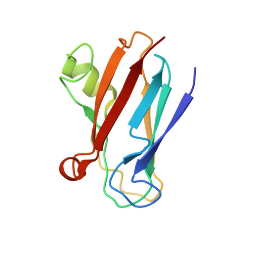
Structure of plastocyanin from the cyanobacterium Anabaena variabilis.

Schmidt, L.Christensen, H.E.Harris, P.

(2006) Acta Crystallogr D Biol Crystallogr 62: 1022-1029

  • DOI: https://doi.org/10.1107/S0907444906023638
  • Primary Citation of Related Structures:  
    2GIM

  • PubMed Abstract: 

    Plastocyanin from the cyanobacterium Anabaena variabilis was heterologously produced in Escherichia coli and purified. Plate-like crystals were obtained by crystallization in 1.15 M trisodium citrate and 7.67 mM sodium borate buffer pH 8.5. The crystals belong to the orthorhombic space group P2(1)2(1)2(1), with unit-cell parameters a = 67.85, b = 45.81, c = 63.41 Angstrom. The structure of the oxidized protein was solved to a resolution of 1.6 Angstrom using plastocyanin from Phormidium laminosum as a search model. Two molecules were found in the asymmetric unit. The electrostatic surface of the basic protein showed a large population of positively charged residues in the northern site, whereas the eastern site lacked the two strongly negatively charged patches. The copper ion was found to be relatively mobile and there were two distinct conformations of His61.


  • Organizational Affiliation

    Department of Chemistry, The Technical University of Denmark, Building 207, DK-2800 Kgs. Lyngby, Denmark.


Macromolecules
Find similar proteins by:  (by identity cutoff)  |  3D Structure
Entity ID: 1
MoleculeChains Sequence LengthOrganismDetailsImage
PlastocyaninA,
B [auth C]
106Trichormus variabilisMutation(s): 0 
Gene Names: petE
UniProt
Find proteins for P0C178 (Anabaena variabilis)
Explore P0C178 
Go to UniProtKB:  P0C178
Entity Groups  
Sequence Clusters30% Identity50% Identity70% Identity90% Identity95% Identity100% Identity
UniProt GroupP0C178
Sequence Annotations
Expand
  • Reference Sequence
Experimental Data & Validation

Experimental Data

  • Method: X-RAY DIFFRACTION
  • Resolution: 1.60 Å
  • R-Value Free: 0.260 
  • R-Value Work: 0.209 
  • R-Value Observed: 0.211 
  • Space Group: P 21 21 21
Unit Cell:
Length ( Å )Angle ( ˚ )
a = 67.85α = 90
b = 45.81β = 90
c = 63.41γ = 90
Software Package:
Software NamePurpose
REFMACrefinement
MAR345data collection
XDSdata scaling
MOLREPphasing

Structure Validation

View Full Validation Report


Entry History 

Deposition Data

Revision History  (Full details and data files)

  • Version 1.0: 2006-04-11
    Type: Initial release
  • Version 1.1: 2008-05-01
    Changes: Version format compliance
  • Version 1.2: 2011-07-13
    Changes: Version format compliance
  • Version 1.3: 2017-10-18
    Changes: Data collection, Refinement description
  • Version 1.4: 2023-08-30
    Changes: Data collection, Database references, Derived calculations, Refinement description