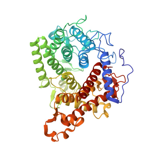
Structure of the complex of a yeast glucoamylase with acarbose reveals the presence of a raw starch binding site on the catalytic domain.
Sevcik, J., Hostinova, E., Solovicova, A., Gasperik, J., Dauter, Z., Wilson, K.S.(2006) FEBS J 273: 2161-2171
- PubMed: 16649993
- DOI: https://doi.org/10.1111/j.1742-4658.2006.05230.x
- Primary Citation of Related Structures:
2F6D, 2FBA - PubMed Abstract:
Most glucoamylases (alpha-1,4-D-glucan glucohydrolase, EC 3.2.1.3) have structures consisting of both a catalytic and a starch binding domain. The structure of a glucoamylase from Saccharomycopsis fibuligera HUT 7212 (Glu), determined a few years ago, consists of a single catalytic domain. The structure of this enzyme with the resolution extended to 1.1 A and that of the enzyme-acarbose complex at 1.6 A resolution are presented here. The structure at atomic resolution, besides its high accuracy, shows clearly the influence of cryo-cooling, which is manifested in shrinkage of the molecule and lowering the volume of the unit cell. In the structure of the complex, two acarbose molecules are bound, one at the active site and the second at a site remote from the active site, curved around Tyr464 which resembles the inhibitor molecule in the 'sugar tongs' surface binding site in the structure of barley alpha-amylase isozyme 1 complexed with a thiomalto-oligosaccharide. Based on the close similarity in sequence of glucoamylase Glu, which does not degrade raw starch, to that of glucoamylase (Glm) from S. fibuligera IFO 0111, a raw starch-degrading enzyme, it is reasonable to expect the presence of the remote starch binding site at structurally equivalent positions in both enzymes. We propose the role of this site is to fix the enzyme onto the surface of a starch granule while the active site degrades the polysaccharide. This hypothesis is verified here by the preparation of mutants of glucoamylases Glu and Glm.
Organizational Affiliation:
Institute of Molecular Biology, Slovak Academy of Sciences, Bratislava, Slovakia. jozef.sevcik@savba.sk