2CJG

Lysine aminotransferase from M. tuberculosis in bound PMP form


Experimental Data Snapshot

  • Method: X-RAY DIFFRACTION
  • Resolution: 1.95 Å
  • R-Value Free: 0.186 
  • R-Value Work: 0.159 
  • R-Value Observed: 0.161 

wwPDB Validation   3D Report Full Report


Ligand Structure Quality Assessment 


This is version 1.3 of the entry. See complete history


Literature

Direct Evidence for a Glutamate Switch Necessary for Substrate Recognition: Crystal Structures of Lysine Epsilon-Aminotransferase (Rv3290C) from Mycobacterium Tuberculosis H37Rv.

Tripathi, S.M.Ramachandran, R.

(2006) J Mol Biol 362: 877

  • DOI: https://doi.org/10.1016/j.jmb.2006.08.019
  • Primary Citation of Related Structures:  
    2CIN, 2CJD, 2CJG, 2CJH

  • PubMed Abstract: 

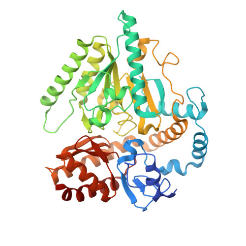
    Lysine epsilon-aminotransferase (LAT) is a PLP-dependent enzyme that is highly up-regulated in nutrient-starved tuberculosis models. It catalyzes an overall reaction involving the transfer of the epsilon-amino group of L-lysine to alpha-ketoglutarate to yield L-glutamate and alpha-aminoadipate-delta-semialdehyde. We have cloned and characterized the enzyme from Mycobacterium tuberculosisH37Rv. We report here the crystal structures of the enzyme, the first from any source, in the unliganded form, external aldimine with L-lysine, with bound PMP and with its C5 substrate alpha-ketoglutarate. In addition to interaction details in the active site, the structures reveal a Glu243 "switch" through which the enzyme changes substrate specificities. The unique substrate L-lysine is recognized specifically when Glu243 maintains a salt-bridge with Arg422. On the other hand, the binding of the common C5 substrates L-glutamate and alpha-ketoglutarate is enabled when Glu243 switches away and unshields Arg422. The structures reported here, sequence conservation and earlier mutational studies suggest that the "glutamate switch" is an elegant solution devised by a subgroup of fold type I aminotransferases for recognition of structurally diverse substrates in the same binding site and provides for reaction specificity.


  • Organizational Affiliation

    Molecular and Structural Biology Division, Central Drug Research Institute, Chattar Manzil, Mahatma Gandhi Marg, Lucknow-226001, India.


Macromolecules
Find similar proteins by:  (by identity cutoff)  |  3D Structure
Entity ID: 1
MoleculeChains Sequence LengthOrganismDetailsImage
L-LYSINE-EPSILON AMINOTRANSFERASE449Mycobacterium tuberculosis H37RvMutation(s): 0 
EC: 2.6.1.36
UniProt
Find proteins for P9WQ77 (Mycobacterium tuberculosis (strain ATCC 25618 / H37Rv))
Explore P9WQ77 
Go to UniProtKB:  P9WQ77
Entity Groups  
Sequence Clusters30% Identity50% Identity70% Identity90% Identity95% Identity100% Identity
UniProt GroupP9WQ77
Sequence Annotations
Expand
  • Reference Sequence
Small Molecules
Ligands 1 Unique
IDChains Name / Formula / InChI Key2D Diagram3D Interactions
PMP
Query on PMP

Download Ideal Coordinates CCD File 
B [auth A]4'-DEOXY-4'-AMINOPYRIDOXAL-5'-PHOSPHATE
C8 H13 N2 O5 P
ZMJGSOSNSPKHNH-UHFFFAOYSA-N
Experimental Data & Validation

Experimental Data

  • Method: X-RAY DIFFRACTION
  • Resolution: 1.95 Å
  • R-Value Free: 0.186 
  • R-Value Work: 0.159 
  • R-Value Observed: 0.161 
  • Space Group: P 31 2 1
Unit Cell:
Length ( Å )Angle ( ˚ )
a = 103.27α = 90
b = 103.27β = 90
c = 98.266γ = 120
Software Package:
Software NamePurpose
REFMACrefinement
MOSFLMdata reduction
SCALAdata scaling
AMoREphasing

Structure Validation

View Full Validation Report


Ligand Structure Quality Assessment 


Entry History 

Deposition Data

Revision History  (Full details and data files)

  • Version 1.0: 2006-08-14
    Type: Initial release
  • Version 1.1: 2011-05-08
    Changes: Version format compliance
  • Version 1.2: 2011-07-13
    Changes: Version format compliance
  • Version 1.3: 2018-01-24
    Changes: Source and taxonomy