2CB8

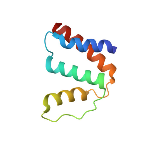
High resolution crystal structure of liganded human L-ACBP


Experimental Data Snapshot

  • Method: X-RAY DIFFRACTION
  • Resolution: 1.40 Å
  • R-Value Free: 0.194 
  • R-Value Work: 0.171 
  • R-Value Observed: 0.172 

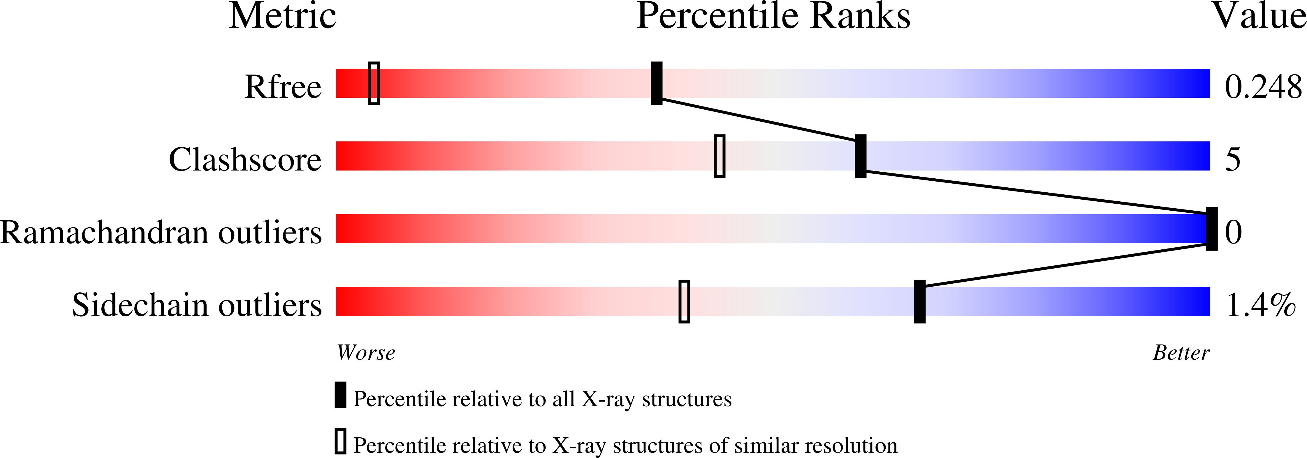
wwPDB Validation   3D Report Full Report


This is version 1.2 of the entry. See complete history


Literature

High Resolution Crystal Structures of Unliganded and Liganded Human Liver Acbp Reveal a New Mode of Binding for the Acyl-Coa Ligand.

Taskinen, J.P.Van Aalten, D.M.Knudsen, J.Wierenga, R.K.

(2007) Proteins 66: 229

  • DOI: https://doi.org/10.1002/prot.21124
  • Primary Citation of Related Structures:  
    2CB8, 2FJ9

  • PubMed Abstract: 

    The acyl-CoA binding protein (ACBP) is essential for the fatty acid metabolism, membrane structure, membrane fusion, and ceramide synthesis. Here high resolution crystal structures of human cytosolic liver ACBP, unliganded and liganded with a physiological ligand, myristoyl-CoA are described. The binding of the acyl-CoA molecule induces only few structural differences near the binding pocket. The crystal form of the liganded ACBP, which has two ACBP molecules in the asymmetric unit, shows that in human ACBP the same acyl-CoA binding pocket is present as previously described for the bovine and Plasmodium falciparum ACBP and the mode of binding of the 3'-phosphate-AMP moiety is conserved. Unexpectedly, in one of the acyl-CoA binding pockets the acyl moiety is bound in a reversed mode as compared with the bovine and P. falciparum structures. In this binding mode, the myristoyl-CoA molecule is fully ordered and bound across the two ACBP molecules of the crystallographic asymmetric unit: the 3'-phosphate-AMP moiety is bound in the binding pocket of one ACBP molecule and the acyl chain is bound in the pocket of the other ACBP molecule. The remaining binding pocket cavities of these two ACBP molecules are filled by other ligand fragments. This novel binding mode shows that the acyl moiety can flip out of its classical binding pocket and bind elsewhere, suggesting a mechanism for the acyl-CoA transfer between ACBP and the active site of a target enzyme. This mechanism is of possible relevance for the in vivo function of ACBP.


  • Organizational Affiliation

    Biocenter Oulu and Department of Biochemistry, University of Oulu, FIN-90014, Finland.


Macromolecules
Find similar proteins by:  (by identity cutoff)  |  3D Structure
Entity ID: 1
MoleculeChains Sequence LengthOrganismDetailsImage
ACYL-COA-BINDING PROTEIN
A, B
87Homo sapiensMutation(s): 0 
UniProt & NIH Common Fund Data Resources
Find proteins for P07108 (Homo sapiens)
Explore P07108 
Go to UniProtKB:  P07108
PHAROS:  P07108
GTEx:  ENSG00000155368 
Entity Groups  
Sequence Clusters30% Identity50% Identity70% Identity90% Identity95% Identity100% Identity
UniProt GroupP07108
Sequence Annotations
Expand
  • Reference Sequence
Small Molecules
Ligands 4 Unique
IDChains Name / Formula / InChI Key2D Diagram3D Interactions
MYA
Query on MYA

Download Ideal Coordinates CCD File 
C [auth A],
I [auth A],
L [auth B],
M [auth B]
TETRADECANOYL-COA
C35 H62 N7 O17 P3 S
DUAFKXOFBZQTQE-QSGBVPJFSA-N
SO4
Query on SO4

Download Ideal Coordinates CCD File 
J [auth A]SULFATE ION
O4 S
QAOWNCQODCNURD-UHFFFAOYSA-L
MXE
Query on MXE

Download Ideal Coordinates CCD File 
P [auth B]2-METHOXYETHANOL
C3 H8 O2
XNWFRZJHXBZDAG-UHFFFAOYSA-N
ZN
Query on ZN

Download Ideal Coordinates CCD File 
D [auth A]
E [auth A]
F [auth A]
G [auth A]
H [auth A]
D [auth A],
E [auth A],
F [auth A],
G [auth A],
H [auth A],
K [auth A],
N [auth B],
O [auth B]
ZINC ION
Zn
PTFCDOFLOPIGGS-UHFFFAOYSA-N
Binding Affinity Annotations 
IDSourceBinding Affinity
MYA Binding MOAD:  2CB8 Kd: 2.5 (nM) from 1 assay(s)
Experimental Data & Validation

Experimental Data

  • Method: X-RAY DIFFRACTION
  • Resolution: 1.40 Å
  • R-Value Free: 0.194 
  • R-Value Work: 0.171 
  • R-Value Observed: 0.172 
  • Space Group: I 2 3
Unit Cell:
Length ( Å )Angle ( ˚ )
a = 118.49α = 90
b = 118.49β = 90
c = 118.49γ = 90
Software Package:
Software NamePurpose
REFMACrefinement
XDSdata reduction
XDSdata scaling
CNSphasing

Structure Validation

View Full Validation Report


Entry History 

Deposition Data

Revision History  (Full details and data files)

  • Version 1.0: 2006-10-25
    Type: Initial release
  • Version 1.1: 2011-05-08
    Changes: Version format compliance
  • Version 1.2: 2011-07-13
    Changes: Version format compliance