2BAP

Crystal structure of the N-terminal mDia1 Armadillo Repeat Region and Dimerisation Domain in complex with the mDia1 autoregulatory domain (DAD)


Experimental Data Snapshot

  • Method: X-RAY DIFFRACTION
  • Resolution: 3.30 Å
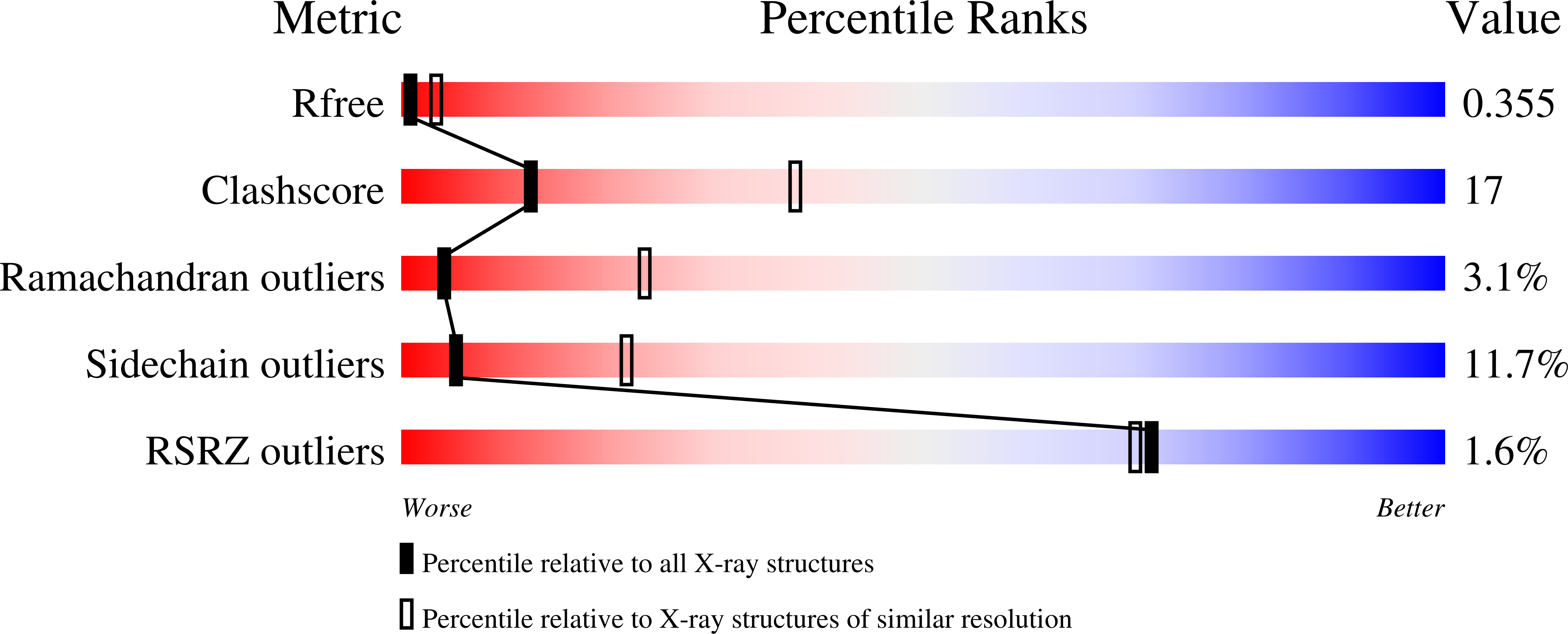
  • R-Value Free: 0.364 
  • R-Value Work: 0.288 
  • R-Value Observed: 0.292 

wwPDB Validation   3D Report Full Report


This is version 1.4 of the entry. See complete history


Literature

The regulation of mDia1 by autoinhibition and its release by Rho*GTP.

Lammers, M.Rose, R.Scrima, A.Wittinghofer, A.

(2005) EMBO J 24: 4176-4187

  • DOI: https://doi.org/10.1038/sj.emboj.7600879
  • Primary Citation of Related Structures:  
    2BAP

  • PubMed Abstract: 

    Formins induce the nucleation and polymerisation of unbranched actin filaments via the formin-homology domains 1 and 2. Diaphanous-related formins (Drfs) are regulated by a RhoGTPase-binding domain situated in the amino-terminal (N-terminal) region and a carboxy-terminal Diaphanous-autoregulatory domain (DAD), whose interaction stabilises an autoinhibited inactive conformation. Binding of active Rho releases DAD and activates the catalytic activity of mDia. Here, we report on the interaction of DAD with the regulatory N-terminus of mDia1 (mDia(N)) and its release by Rho*GTP. We have defined the elements required for tight binding and solved the three-dimensional structure of a complex between an mDia(N) construct and DAD by X-ray crystallography. The core DAD region is an alpha-helical peptide, which binds in the most highly conserved region of mDia(N) using mainly hydrophobic interactions. The structure suggests a two-step mechanism for release of autoinhibition whereby Rho*GTP, although having a partially nonoverlapping binding site, displaces DAD by ionic repulsion and steric clashes. We show that Rho*GTP accelerates the dissociation of DAD from the mDia(N)*DAD complex.


  • Organizational Affiliation

    Department of Structural Biology, Max-Planck-Institute of Molecular Physiology, Dortmund, Germany.


Macromolecules
Find similar proteins by:  (by identity cutoff)  |  3D Structure
Entity ID: 1
MoleculeChains Sequence LengthOrganismDetailsImage
Diaphanous protein homolog 1A [auth B],
B [auth A]
317Mus musculusMutation(s): 0 
Gene Names: Diaph1Diap1
UniProt & NIH Common Fund Data Resources
Find proteins for O08808 (Mus musculus)
Explore O08808 
Go to UniProtKB:  O08808
IMPC:  MGI:1194490
Entity Groups  
Sequence Clusters30% Identity50% Identity70% Identity90% Identity95% Identity100% Identity
UniProt GroupO08808
Sequence Annotations
Expand
  • Reference Sequence
Find similar proteins by:  (by identity cutoff)  |  3D Structure
Entity ID: 2
MoleculeChains Sequence LengthOrganismDetailsImage
Diaphanous protein homolog 1C [auth D],
D [auth C]
56Mus musculusMutation(s): 0 
Gene Names: Diaph1Diap1
UniProt & NIH Common Fund Data Resources
Find proteins for O08808 (Mus musculus)
Explore O08808 
Go to UniProtKB:  O08808
IMPC:  MGI:1194490
Entity Groups  
Sequence Clusters30% Identity50% Identity70% Identity90% Identity95% Identity100% Identity
UniProt GroupO08808
Sequence Annotations
Expand
  • Reference Sequence
Experimental Data & Validation

Experimental Data

  • Method: X-RAY DIFFRACTION
  • Resolution: 3.30 Å
  • R-Value Free: 0.364 
  • R-Value Work: 0.288 
  • R-Value Observed: 0.292 
  • Space Group: P 61 2 2
Unit Cell:
Length ( Å )Angle ( ˚ )
a = 138.45α = 90
b = 138.45β = 90
c = 210.89γ = 120
Software Package:
Software NamePurpose
XSCALEdata scaling
PHASERphasing
REFMACrefinement
PDB_EXTRACTdata extraction
MAR345data collection

Structure Validation

View Full Validation Report


Entry History 

Deposition Data

Revision History  (Full details and data files)

  • Version 1.0: 2006-03-07
    Type: Initial release
  • Version 1.1: 2008-05-01
    Changes: Version format compliance
  • Version 1.2: 2011-07-13
    Changes: Version format compliance
  • Version 1.3: 2017-10-11
    Changes: Refinement description
  • Version 1.4: 2023-08-23
    Changes: Data collection, Database references, Refinement description