2ANJ

Crystal Structure of the Glur2 Ligand Binding Core (S1S2J-Y450W) Mutant in Complex With the Partial Agonist Kainic Acid at 2.1 A Resolution


Experimental Data Snapshot

  • Method: X-RAY DIFFRACTION
  • Resolution: 2.10 Å
  • R-Value Free: 0.248 
  • R-Value Work: 0.187 
  • R-Value Observed: 0.187 

wwPDB Validation   3D Report Full Report


Ligand Structure Quality Assessment 


This is version 1.6 of the entry. See complete history


Literature

A Binding Site Tyrosine Shapes Desensitization Kinetics and Agonist Potency at GluR2: a mutagenic, kinetic, and crystallographic study

Holm, M.M.Naur, P.Vestergaard, B.Geballe, M.T.Gajhede, M.Kastrup, J.S.Traynelis, S.F.Egebjerg, J.

(2005) J Biol Chem 280: 35469-35476

  • DOI: https://doi.org/10.1074/jbc.M507800200
  • Primary Citation of Related Structures:  
    2ANJ

  • PubMed Abstract: 

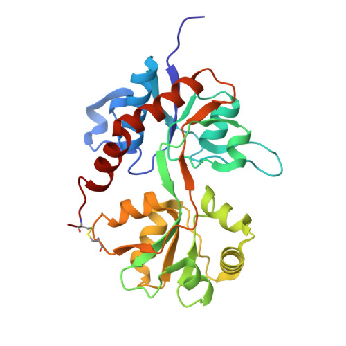
    Binding of an agonist to the 2-amino-3-(3-hydroxy-5-methyl-4-isoxazolyl)-propionic acid (AMPA) receptor family of the glutamate receptors (GluRs) results in rapid activation of an ion channel. Continuous application results in a non-desensitizing response for agonists like kainate, whereas most other agonists, such as the endogenous agonist (S)-glutamate, induce desensitization. We demonstrate that a highly conserved tyrosine, forming a wedge between the agonist and the N-terminal part of the bi-lobed ligand-binding site, plays a key role in the receptor kinetics as well as agonist potency and selectivity. The AMPA receptor GluR2, with mutations in Tyr-450, were expressed in Xenopus laevis oocytes and characterized in a two-electrode voltage clamp setup. The mutation GluR2(Y450A) renders the receptor highly kainate selective, and rapid application of kainate to outside-out patches induced strongly desensitizing currents. When Tyr-450 was substituted with the larger tryptophan, the (S)-glutamate desensitization is attenuated with a 10-fold increase in steady-state/peak currents (19% compared with 1.9% at the wild type). Furthermore, the tryptophan mutant was introduced into the GluR2-S1S2J ligand binding core construct and co-crystallized with kainate, and the 2.1-A x-ray structure revealed a slightly more closed ligand binding core as compared with the wild-type complex. Through genetic manipulations combined with structural and electrophysiological analysis, we report that mutations in position 450 invert the potency of two central agonists while concurrently strongly shaping the agonist efficacy and the desensitization kinetics of the AMPA receptor GluR2.


  • Organizational Affiliation

    Department of Molecular Biology, C. F. Møllers Allé Bldg. 130, University of Aarhus, DK-8000 Aarhus, Denmark. mmh@fi.au.dk


Macromolecules
Find similar proteins by:  (by identity cutoff)  |  3D Structure
Entity ID: 1
MoleculeChains Sequence LengthOrganismDetailsImage
Glutamate receptor 2263Rattus norvegicusMutation(s): 1 
Gene Names: Gria2Glur2
UniProt
Find proteins for P19491 (Rattus norvegicus)
Explore P19491 
Go to UniProtKB:  P19491
Entity Groups  
Sequence Clusters30% Identity50% Identity70% Identity90% Identity95% Identity100% Identity
UniProt GroupP19491
Sequence Annotations
Expand
  • Reference Sequence
Small Molecules
Ligands 1 Unique
IDChains Name / Formula / InChI Key2D Diagram3D Interactions
KAI
Query on KAI

Download Ideal Coordinates CCD File 
B [auth A]3-(CARBOXYMETHYL)-4-ISOPROPENYLPROLINE
C10 H15 N O4
VLSMHEGGTFMBBZ-OOZYFLPDSA-N
Binding Affinity Annotations 
IDSourceBinding Affinity
KAI BindingDB:  2ANJ Ki: min: 1.8, max: 1.22e+4 (nM) from 11 assay(s)
EC50: min: 1.06e+4, max: 3.80e+5 (nM) from 8 assay(s)
Experimental Data & Validation

Experimental Data

  • Method: X-RAY DIFFRACTION
  • Resolution: 2.10 Å
  • R-Value Free: 0.248 
  • R-Value Work: 0.187 
  • R-Value Observed: 0.187 
  • Space Group: P 21 21 2
Unit Cell:
Length ( Å )Angle ( ˚ )
a = 95.254α = 90
b = 61.635β = 90
c = 48.751γ = 90
Software Package:
Software NamePurpose
DENZOdata reduction
SCALEPACKdata scaling
AMoREphasing
CNSrefinement
PDB_EXTRACTdata extraction

Structure Validation

View Full Validation Report


Ligand Structure Quality Assessment 


Entry History 

Deposition Data

Revision History  (Full details and data files)

  • Version 1.0: 2005-08-30
    Type: Initial release
  • Version 1.1: 2008-04-01
    Changes: Version format compliance
  • Version 1.2: 2011-07-13
    Changes: Version format compliance
  • Version 1.3: 2017-08-02
    Changes: Data collection, Refinement description, Source and taxonomy
  • Version 1.4: 2017-10-11
    Changes: Refinement description
  • Version 1.5: 2021-10-20
    Changes: Database references, Derived calculations
  • Version 1.6: 2023-08-23
    Changes: Data collection, Refinement description