2ERV

Crystal structure of the outer membrane enzyme PagL


Experimental Data Snapshot

  • Method: X-RAY DIFFRACTION
  • Resolution: 2.00 Å
  • R-Value Free: 0.233 
  • R-Value Work: 0.198 
  • R-Value Observed: 0.200 

wwPDB Validation   3D Report Full Report


Ligand Structure Quality Assessment 


This is version 1.4 of the entry. See complete history


Literature

Crystal structure and catalytic mechanism of the LPS 3-O-deacylase PagL from Pseudomonas aeruginosa.

Rutten, L.Geurtsen, J.Lambert, W.Smolenaers, J.J.Bonvin, A.M.de Haan, A.van der Ley, P.Egmond, M.R.Gros, P.Tommassen, J.

(2006) Proc Natl Acad Sci U S A 103: 7071-7076

  • DOI: https://doi.org/10.1073/pnas.0509392103
  • Primary Citation of Related Structures:  
    2ERV

  • PubMed Abstract: 

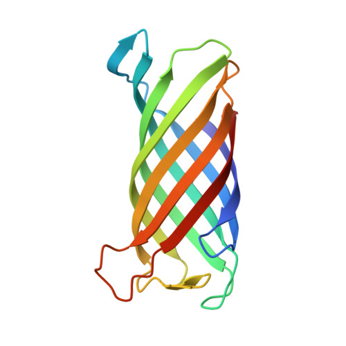
    Pathogenic gram-negative bacteria can modify the lipid A portion of their lipopolysaccharide in response to environmental stimuli. 3-O-deacylation of lipid A by the outer membrane enzyme PagL modulates signaling through Toll-like receptor 4, leading to a reduced host immune response. We found that PagL is widely disseminated among gram-negative bacteria. Only four residues are conserved: a Ser, His, Phe, and Asn residue. Here, we describe the crystal structure of PagL from Pseudomonas aeruginosa to 2.0-A resolution. It consists of an eight-stranded beta-barrel with the axis tilted by approximately 30 degrees with respect to the lipid bilayer. The structure reveals that PagL contains an active site with a Ser-His-Glu catalytic triad and an oxyanion hole that comprises the conserved Asn. The importance of active site residues was confirmed in mutagenesis studies. Although PagL is most likely active as a monomer, its active site architecture shows high resemblance to that of the dimeric 12-stranded outer membrane phospholipase A. Modeling of the substrate lipid X onto the active site reveals that the 3-O-acyl chain is accommodated in a hydrophobic groove perpendicular to the membrane plane. In addition, an aspartate makes a hydrogen bond with the hydroxyl group of the 3-O-acyl chain, probably providing specificity of PagL toward lipid A.


  • Organizational Affiliation

    Department of Crystal and Structural Chemistry, Bijvoet Center for Biomolecular Research, Utrecht, The Netherlands.


Macromolecules
Find similar proteins by:  (by identity cutoff)  |  3D Structure
Entity ID: 1
MoleculeChains Sequence LengthOrganismDetailsImage
hypothetical protein Paer03002360
A, B
150Pseudomonas aeruginosaMutation(s): 0 
Gene Names: PA4661
Membrane Entity: Yes 
UniProt
Find proteins for Q9HVD1 (Pseudomonas aeruginosa (strain ATCC 15692 / DSM 22644 / CIP 104116 / JCM 14847 / LMG 12228 / 1C / PRS 101 / PAO1))
Explore Q9HVD1 
Go to UniProtKB:  Q9HVD1
Entity Groups  
Sequence Clusters30% Identity50% Identity70% Identity90% Identity95% Identity100% Identity
UniProt GroupQ9HVD1
Sequence Annotations
Expand
  • Reference Sequence
Small Molecules
Ligands 2 Unique
IDChains Name / Formula / InChI Key2D Diagram3D Interactions
CXE
Query on CXE

Download Ideal Coordinates CCD File 
D [auth A]
E [auth A]
F [auth A]
G [auth A]
H [auth B]
D [auth A],
E [auth A],
F [auth A],
G [auth A],
H [auth B],
I [auth B],
J [auth B],
K [auth B],
L [auth B],
M [auth B],
N [auth B],
O [auth B]
PENTAETHYLENE GLYCOL MONODECYL ETHER
C20 H42 O6
QAXPOSPGRHYIHE-UHFFFAOYSA-N
CA
Query on CA

Download Ideal Coordinates CCD File 
C [auth A]CALCIUM ION
Ca
BHPQYMZQTOCNFJ-UHFFFAOYSA-N
Experimental Data & Validation

Experimental Data

  • Method: X-RAY DIFFRACTION
  • Resolution: 2.00 Å
  • R-Value Free: 0.233 
  • R-Value Work: 0.198 
  • R-Value Observed: 0.200 
  • Space Group: C 1 2 1
Unit Cell:
Length ( Å )Angle ( ˚ )
a = 92.264α = 90
b = 48.993β = 115.46
c = 105.026γ = 90
Software Package:
Software NamePurpose
DENZOdata reduction
SCALEPACKdata scaling
PHASERphasing
RESOLVEphasing
REFMACrefinement
PDB_EXTRACTdata extraction

Structure Validation

View Full Validation Report


Ligand Structure Quality Assessment 


Entry History 

Deposition Data

Revision History  (Full details and data files)

  • Version 1.0: 2006-04-11
    Type: Initial release
  • Version 1.1: 2008-05-01
    Changes: Version format compliance
  • Version 1.2: 2011-07-13
    Changes: Advisory, Version format compliance
  • Version 1.3: 2017-10-18
    Changes: Advisory, Refinement description
  • Version 1.4: 2023-08-23
    Changes: Advisory, Data collection, Database references, Derived calculations, Refinement description